the Creative Commons Attribution 4.0 License.
the Creative Commons Attribution 4.0 License.
 | 14 Oct 2026
The co-occurrence of peri-prosthetic fracture and infection: a scoping review on current diagnostic principles, treatment and outcome
Noémie Reinert
Laura Bessems
Martin Clauss
Nader Maai
Jolien Onsea
Georges Vles
Karl Stoffel
Willem-Jan Metsemakers
Mario Morgenstern

Background: The co-occurrence of periprosthetic fractures and infection, referred to as infected periprosthetic fracture (iPPF), represents a highly burdensome complication in orthopedic surgery. Management is challenging since not rarely treatment principles of prosthetic joint infections and fracture-related infections have to be combined. The aim of this scoping review was to map the current literature on management and outcome of iPPFs. Methods: A scoping review was conducted in accordance with PRISMA-ScR guidelines. Five major databases were systematically searched. Studies reporting on diagnostic principles and/or treatment strategies for iPPF around the hip and knee were included. Data were mapped regarding study design, diagnostic approaches, surgical strategies, and outcome. Results: A total of 32 studies comprising 273 patients with iPPFs were included; 8 addressed diagnostic strategies, and 24 reported on treatment and outcome. Diagnostic studies demonstrated limited reliability of serum inflammatory markers, and although synovial fluid analysis appeared most informative, optimal thresholds remain uncertain. Most treatment studies were small and heterogeneous. A staged surgical approach, predominantly two-stage revision, was reported in 83 % of the included studies. Across nine studies reporting outcomes, mean infection eradication and recurrence rates were 76.1 % and 22.3 %, respectively. The majority of the studies (58.3 %) described that additional surgeries were needed to cure infection. Fracture healing outcomes were inconsistently reported (bone healing in 4 out of 9 studies with 53/58 fractures, mean 91.4 %). Conclusion: This scoping review demonstrates that management of iPPFs remains highly challenging, with high rates of recurrent infection and unplanned revision surgery. However, the reported quantitative outcome data represent descriptive summaries derived from heterogeneous, predominantly small observational studies and case series and should not be interpreted as definitive benchmarks for clinical outcomes. The current evidence base remains limited, highlighting the need for standardized diagnostic criteria, uniform outcome reporting, and well-designed prospective multicenter studies.
- Article
(711 KB) - Full-text XML
- BibTeX
- EndNote


Periprosthetic fractures (PPFs) are known to be devastating complications after joint arthroplasty and are associated with considerable morbidity and mortality. With an aging population and a continuous rise in arthroplasty procedures, the incidence of PPFs is increasing and represents a growing clinical and socioeconomic challenge (Rupp et al., 2022). These injuries are associated with high complication rates such as instability, nonunion or infection (El Khassawna et al., 2021; Moreta et al., 2021), and high mortality rate up to 60 % (Walter et al., 2024). In the recent literature, high infection rates ranging from 5.6 % to 16.1 % for PPFs have been reported (Luyckx et al., 2022; Miettinen et al., 2023; Preston et al., 2015). Notably, even infection without fracture (isolated prosthetic joint infection, PJI) carries substantial mortality – 11 % after infected total hip arthroplasty (THA) and 13 % after infected total knee arthroplasty (TKA) (Ramos et al., 2025, 2026), which is comparable to common cancers. The co-occurrence of both PPF and an infection, hereafter referred to as an infected periprosthetic fracture (iPPF), represents a hybrid entity combining aspects of both PJIs and fracture-related infections (FRIs). Consequently, iPPFs require clinicians to be familiar with principles of both PJI and FRI management. However, these objectives may conflict. In chronic PJIs, treatment often involves a two-stage procedure with an antibiotic-loaded spacer and an implant-free interval. In contrast, successful FRI management requires stable fixation to promote bone healing and infection eradication. The management of iPPFs therefore necessitates reconciling two potentially contradictory principles: infection eradication through implant removal and maintenance of mechanical stability (Müller et al., 2019). The diagnosis of iPPF is equally demanding, since inflammatory markers are not reliable and of poor predictive value in the history of recent trauma (Chevillotte et al., 2009).
Since existing evidence of iPPFs is sparse and heterogeneous, the objective of this scoping review is twofold: (1) to systematically map the published treatment concepts and reported outcomes for iPPFs and (2) to provide an overview of current diagnostic principles.
2.1 Reporting guidelines
This scoping review was conducted in accordance with the PRISMA-ScR guidelines (Tricco et al., 2018), and the protocol has been pre-registered on the Open Science Framework platform (Reinert et al., 2025b).
2.2 Literature search strategy
A comprehensive search of the biomedical databases – PubMed (including MEDLINE), Embase, Web of Science Core Collection, Scopus, and CENTRAL (via Cochrane Library) – was conducted on 8 January 2026. The strategy was developed in collaboration with biomedical reference librarians. The search strategy combined MeSH/Emtree terms and free-text keywords representing the concepts “periprosthetic fracture” and “infection”. After finalizing the search strategy in Pubmed, the framework was extended to the other included databases and checked for consistency. Search limits were restricted to the English language. Gray literature was taken into account for a more continuous screening process with the inclusion of Clinicaltrials.gov and the International Clinical Trials Registry Platform (ICTRP).
2.3 Study eligibility and selection
All retrieved records were imported into Endnote Version 21 (Clarivate, USA), and deduplication was performed. Using the Rayyan platform, the primary studies were screened by two independent reviewers (Noemie Reinert and Laura Bessems). In the case of disagreement, a third reviewer was consulted (JO). Titles and abstracts were screened with respect to the predefined eligibility criteria. The full text of the relevant articles was then matched against our inclusion and exclusion criteria, and any reasons for in- or exclusion were documented.
The Joanna Briggs Institute PCC framework for scoping reviews was applied (Pollock et al., 2023):
-
population – patients with a periprosthetic fracture and infection;
-
concept – treatment algorithm and outcome, diagnostic principles;
-
context – hip joint (acetabulum, proximal femur, shaft, distal femur) and knee joint (proximal tibia and shaft).
Eligible study designs included observational studies and case reports. Exclusion criteria were other anatomical locations than hip and knee joint, animal studies, conference abstracts, narrative reviews without original patient data, editorials/letters, and fractures due to malignant/benign tumors or metabolic bone diseases.
2.4 Data extraction and presentation
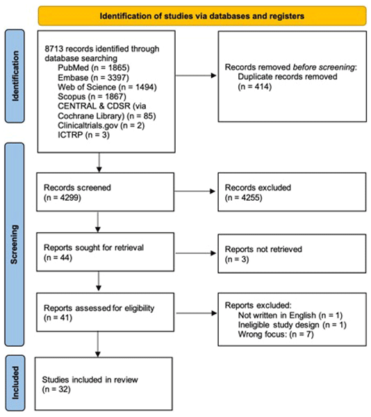
The study selection process was summarized using a PRISMA flow diagram (Fig. 1). Included studies were grouped into diagnostic and treatment papers. Extraction of relevant data was recorded for the following key information: author, title and year of the publication, the study design and sample size, fracture classification, infection definition, surgical treatment concepts addressing iPPF, follow-up intervals, infection outcome, and bone healing. For outcome analysis only studies that include a minimum of five patients were considered.
2.5 Infection scenarios
IPPFs were categorized into two predefined scenarios.
-
Scenario F-I. A PPF is treated with fracture fixation or revision arthroplasty, followed by an infection. Revision arthroplasty is defined by removing and replacing or exchanging one or more prosthetic components due to complication (fracture), e.g. femoral stem revision in a Vancouver B2 fracture (Liebs et al., 2015).
-
Scenario I-F. A PPF occurs in the setting of an already infected prosthesis. This includes fractures as a result of implant loosening with proof of infection in the PPF revision surgery (scenario I-F-A) or fractures during treatment of a confirmed PJI (during explantation, re-implantation, exchange, or between stages) (scenario I-F-B).
After applying the predefined inclusion and exclusion criteria to 4299 retrieved articles, a total of 32 studies reporting on iPPFs were included for analysis and synthesis (Fig. 1). The eligible articles comprising in total 273 patients included 8 diagnostic papers (Table 1) and 24 treatment papers (Table 2).
Table 1Overview of diagnostic papers (n=8).
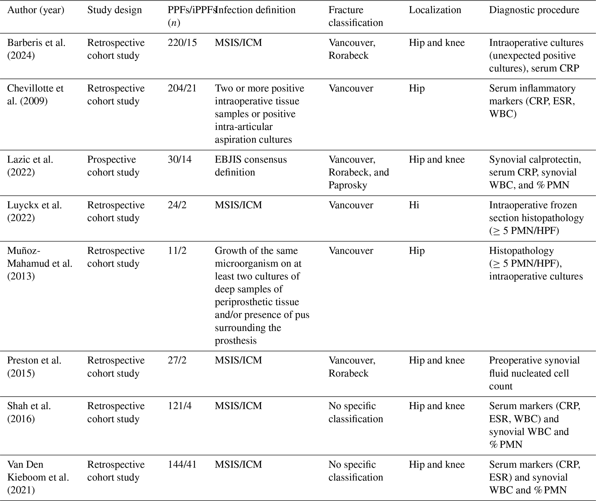
MSIS/ICM: Musculoskeletal Infection Society/International Consensus Meeting; CRP: C-reactive protein; ESR: erythrocyte sedimentation rate; WBC: white blood cell count; EBJIS: European Bone and Joint Infection Society; PMN/HPF: polymorphonuclear cells per high-power field.
Table 2Overview of therapeutic papers (n=24).
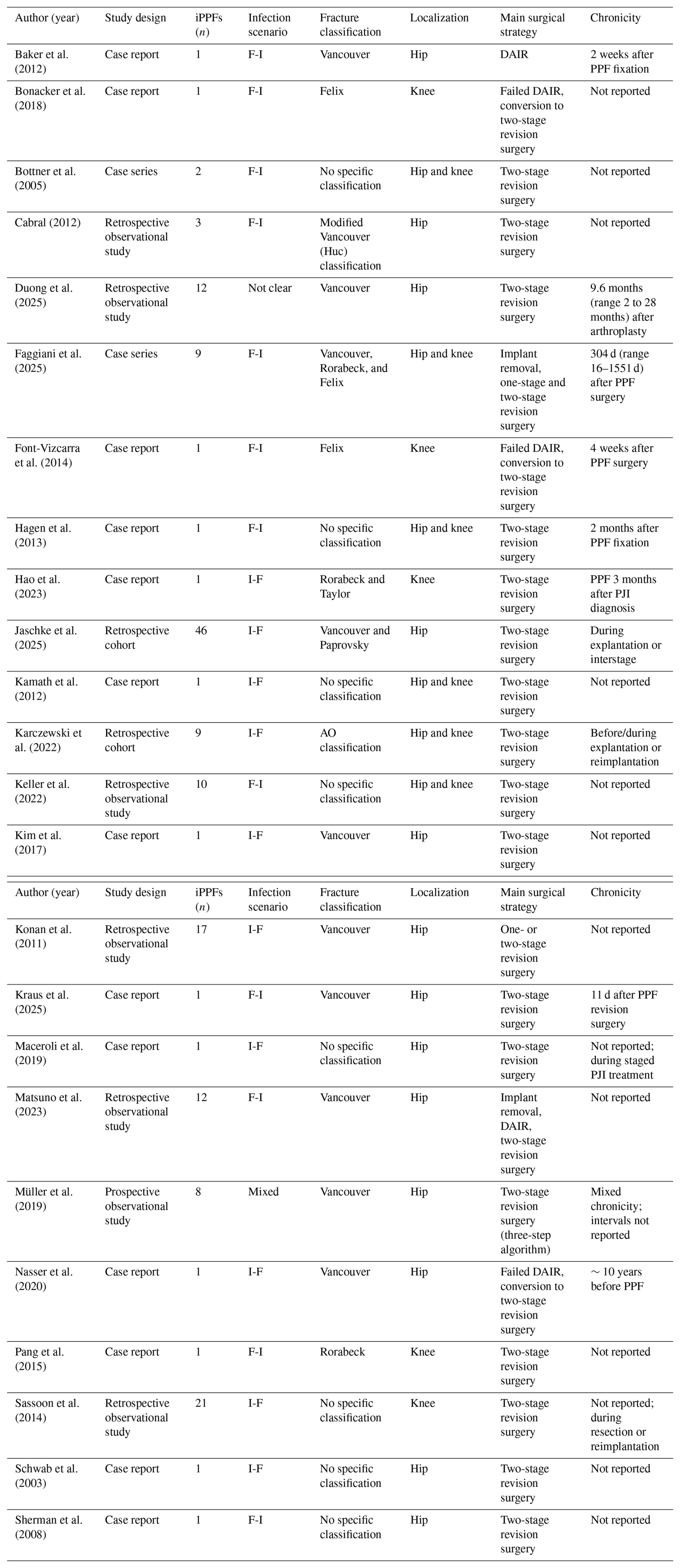
I: infection; F: fracture; PPF: periprosthetic fracture; PJI: periprosthetic joint infection. For F-I cases, timing refers to the interval between PPF treatment and infection onset or diagnosis. For I-F cases, the chronicity of the pre-existing infection and/or the timing of fracture occurrence within the PJI treatment pathway is reported.
3.1 Diagnostic papers
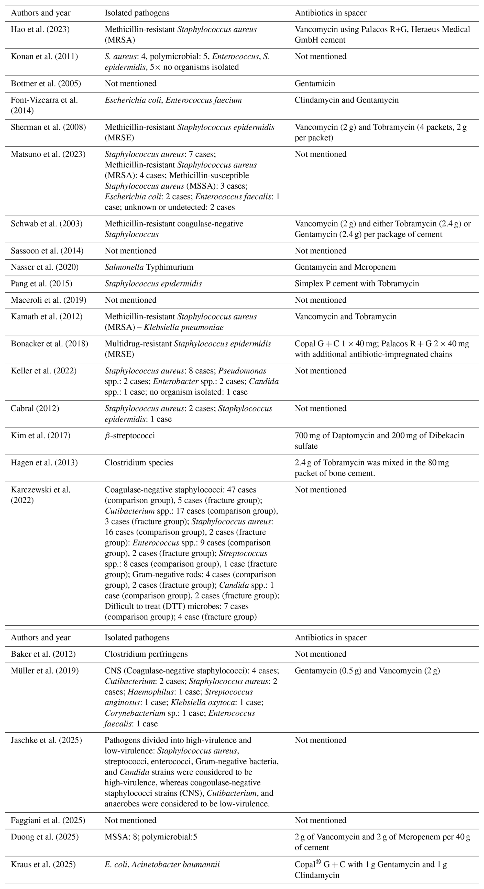
The 8 studies included 111 patients with an iPPF and 670 patients with a PPF but no infection (control group). Across included studies, diagnostic evaluation for concomitant infection employed multiple approaches: serum inflammatory markers (C-reactive protein in eight studies), synovial fluid analysis including nucleated cell counts (four studies), intraoperative frozen section histopathology (two studies), and microbiological culture (standard in all studies) (Table 1) (Barberis et al., 2024; Chevillotte et al., 2009; Lazic et al., 2022; Luyckx et al., 2022; Muñoz-Mahamud et al., 2013; Preston et al., 2015; Shah et al., 2016; Van Den Kieboom et al., 2021). In six of the eight studies, infection was defined according to established criteria: five studies applied the Musculoskeletal Infection Society/International Consensus Meeting (MSIS/ICM) definition (Parvizi and Gehrke, 2014), and one study used the European Bone and Joint Infection Society (EBJIS) definition (McNally et al., 2021). In two studies no specific definition was mentioned. The Vancouver classification was the most commonly used fracture classification system (six out of eight studies), followed by the Rorabeck classification (Rorabeck and Taylor, 1999; Schopper et al., 2022). Data on the retrieved pathogens and used local antibiotics are summarized in Appendix A.
3.2 Treatment papers
An overview of the included 24 treatment papers comprising 162 iPPF cases is presented in Table 2 (Baker et al., 2012; Bonacker et al., 2018; Bottner et al., 2005; Cabral, 2012; Duong et al., 2025; Faggiani et al., 2025; Font-Vizcarra et al., 2014; Hagen et al., 2013; Hao et al., 2023; Jaschke et al., 2025; Kamath et al., 2012; Karczewski et al., 2022; Keller et al., 2022; Kim et al., 2017; Konan et al., 2011; Kraus et al., 2025; Maceroli et al., 2019; Matsuno et al., 2023; Müller et al., 2019; Nasser et al., 2020; Pang et al., 2015; Sassoon et al., 2014; Schwab et al., 2003; Sherman et al., 2008).
In the majority of the included studies (83.3 %; 20/24 with 125 cases) a two-stage procedure was described to treat the iPPF. One-stage exchange was only mentioned in 2 studies including 14 cases (Faggiani et al., 2025; Konan et al., 2011). In two studies a DAIR procedure was applied (Baker et al., 2012; Matsuno et al., 2023), and in three studies (each including three cases) a conversion from a DAIR to a two-stage revision was necessary due to treatment failure (Bonacker et al., 2018; Font-Vizcarra et al., 2014; Nasser et al., 2020). The majority of the studies (58.3 %; 14/24) reported that additional unplanned surgical procedures, beyond the initially intended treatment strategy (DAIR, one-stage exchange, or two-stage exchange), were required to achieve infection eradication.
Outcomes were analyzed across 9 studies, including a total of 146 cases (Table 3) (Duong et al., 2025; Faggiani et al., 2025; Jaschke et al., 2025; Karczewski et al., 2022; Keller et al., 2022; Konan et al., 2011; Matsuno et al., 2023; Müller et al., 2019; Sassoon et al., 2014). Infection eradication rate ranged from 58.3 % to 91.7 %, with a mean infection eradication rate of 76.1 % (99/130). Infection recurrence rate ranged from 8.3 % to 29.2 %, with a mean recurrence rate of 22.3 % (29/130). Bone healing outcome was reported only by 4 studies including 58 cases and ranged from 62.5 % to 100 % (mean 91.4 %; 53/58).
Table 3Infection and radiological outcomes in selected treatment studies (n=9).
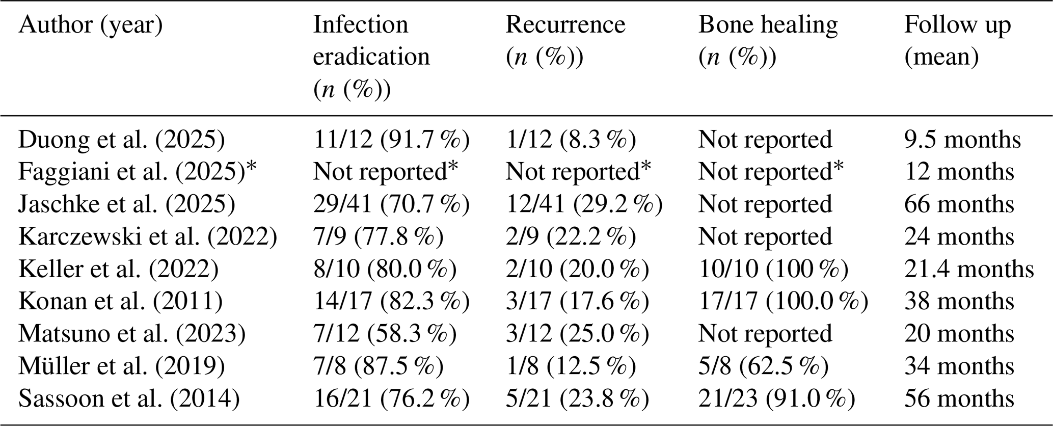
* Included for provided data on follow-up and (additional) surgeries.
IPPFs should be regarded as a hybrid clinical entity that combines characteristics and principles of FRIs and PJIs, and therefore their management must simultaneously address infection eradication, implant stability, and fracture healing.
This scoping review, which includes 32 studies (8 diagnostic and 24 treatment studies) with 273 patients with an iPPF in total, reveals that current evidence is mainly based on small-scale studies and case reports. Evidence of diagnostic strategies is still scarce, and no standardized treatment algorithm exists. Correspondingly, there is limited outcome reporting for iPPFs: infection outcomes were reported for only 130 cases and bone healing for solely 58 cases.
4.1 Diagnostic challenges in iPPFs
Although infection occurs in a relatively high proportion of PPFs, its diagnosis remains challenging as fracture-related inflammation interferes with the accuracy of standard diagnostic tests, and, to date, the current PJI definitions are not validated in the setting of iPPF (McNally et al., 2021). In addition, the reviewed literature demonstrates considerable heterogeneity in the infection definitions. Most included studies applied diagnostic criteria developed for PJI, particularly the MSIS/ICM or EBJIS definitions, whereas the FRI consensus definition was rarely applied, likely reflecting the historical predominance of arthroplasty-focused research. However, as iPPFs represent a hybrid pathology, neither the PJI nor the FRI diagnostic framework alone adequately captures the full spectrum of disease. A diagnostic strategy applying established PJI and FRI definitions in parallel should therefore be adopted, whereas joint involvement determines which diagnostic pathway is most relevant. Since infection may involve the prosthetic joint, the fracture site, or both simultaneously, integrating both diagnostic frameworks enables a more comprehensive assessment of infection extent and provides a stronger basis for treatment planning.
The reviewed studies consistently demonstrate considerable diagnostic limitations and underline that no single parameter is sufficiently reliable on its own. Therefore, the interpretation of laboratory, synovial, and intraoperative findings must always account for the coexistence of trauma (Chevillotte et al., 2009; Luyckx et al., 2022; Muñoz-Mahamud et al., 2013; Shah et al., 2016; Van Den Kieboom et al., 2021).
Chevillotte et al. (2009) illustrated this problem clearly. Although inflammatory markers were frequently elevated in patients with PPFs, their positive predictive value for infection was poor (29 % for CRP, 21 % for ESR, and 18 % for WBC), warranting cautious interpretation of these parameters when a PPF and infection coexist. The high sensitivity but low specificity in this context has also been shown by Van Den Kieboom et al. (2021) when applying MSIS thresholds, promoting combined inflammatory markers to improve the diagnostic performance in iPPFs (Van Den Kieboom et al., 2021). These results demonstrate that serum markers cannot reliably distinguish infection from trauma-related inflammatory response. This is in line with current evidence in FRIs and PJIs. A meta-analysis of serum inflammatory markers in FRIs showed that inflammatory markers are insufficiently accurate to diagnose late FRI (Van Den Kieboom et al., 2018). These results were confirmed for PJIs by Sigmund et al. (2020). In summary serum inflammatory markers show limited diagnostic accuracy in iPPF for two opposite, timing-dependent reasons. Acutely, fracture and surgical trauma themselves trigger a systemic acute-phase response, elevating CRP, ESR, and WBC independent of infection. This is consistent with the observation of Zagra et al. (2026) that serum markers cannot reliably distinguish infection from trauma-related inflammation (Zagra et al., 2026). In late or chronic FRI, the problem shifts to sensitivity: low-grade, biofilm-associated infections caused by low-virulence organisms (e.g., coagulase-negative staphylococci) provoke only a weak systemic response, so markers may remain normal despite active infection – consistent with the FRI consensus definition, which treats serum markers as merely suggestive rather than confirmatory (Metsemakers et al., 2018). Serum markers are thus compromised in opposite directions depending on timing, reinforcing the preference for synovial fluid and microbiological criteria in diagnosing iPPF.
The diagnostic utility of synovial fluid WBC in the subgroup of iPPFs was analyzed in detail by Shah et al. (2016). Although their concern that fracture-related inflammation or hemarthrosis could falsely elevate these values, their results could demonstrate that synovial WBC count provides excellent sensitivity for diagnosing iPPF (with an overall area under the curve (AUC) of 84 %). They identified optimal cut-offs of 2707 WBC µL−1 and 77 % polymorphonuclear cells (PMN), similar to the cut-offs of 3000 WBC µL−1 and 80 % for the % PMNs used in the diagnostic workup of PJI (McNally et al., 2021). In contrast to this, Van Den Kieboom et al. (2021) identified substantially higher cut-off values (4552 WBC µL−1, 79.5 % PMN) compared to PJI alone, suggesting higher thresholds and utilizing all serum and synovial markers in combination (Van Den Kieboom et al., 2021). Preston et al. (2015) analyzed preoperative synovial fluid nucleated cell count and found a positive predictive value (PPV) of only 18 %, indicating that fewer than one-fifth of patients with elevated cell count following fracture ultimately develop iPPF. However, with a sensitivity of 100 %, a normal nucleated cell count effectively excludes infection in PPFs. These tests offer several advantages, including rapid availability, low cost, objectivity, and the ability to be performed pre- or intraoperatively without specialized resources. However, in infected fractures without joint involvement, synovial markers may not be elevated as the joint itself is not affected.
Lazic et al. (2022) evaluated the reliability of calprotectin in the pre- or intraoperative diagnosis of iPPF (Lazic et al., 2022). Calprotectin demonstrated higher sensitivity than synovial WBC and PMN, while WBC and PMN showed high specificity. However, these results may be subject to bias as standard synovial analysis was not feasible in 10 cases, which may lead to an underestimation of the sensitivity of WBC and PMN in their study, involving only 14 cases with confirmed infection.
Two studies analyzed histopathological assessment, including intraoperative frozen sections (Luyckx et al., 2022; Muñoz-Mahamud et al., 2013). While, intraoperative frozen section histopathology is reliable for PJI diagnosis in non-traumatic revisions, its accuracy decreases markedly after a recent fracture due to trauma-induced PMN infiltration. Muñoz-Mahamud et al. (2013) found a 66.6 % false-positive rate for histology results when diagnosing infection in revision arthroplasty for Vancouver B2 periprosthetic fractures (100 % sensitivity, 55 % specificity) (Muñoz-Mahamud et al., 2013). In the study of Luyckx et al. (2022) the PPV was only 5.3 %. In the non-traumatic group, PPV was 80.8 %, with a negative predictive value (NPV) of 95.3 %. The authors suggested to set higher PMN cell count thresholds in the presence of recent trauma, although no specific cut-off values were proposed (Luyckx et al., 2022). This reinforces the notion that histopathology should be interpreted as a supportive, not decisive, diagnostic criterion in iPPFs.
Finally, Barberis et al. (2024) investigated the relevance of unexpected positive cultures. They found positive cultures in 6.8 % of the cases (15/159), while in seven patients two positive cultures were present for the same pathogens, meeting the ICM criteria for PJI. The authors also found a significant association of preoperative elevated CRP in culture-positive PPFs, although this must be considered in light of the poor specificity of CRP in the context of trauma (Barberis et al., 2024).
The reviewed diagnostic studies demonstrate that diagnosing infection in iPPFs relies on careful integration of clinical, laboratory, and intraoperative findings rather than on isolated parameters. Serum inflammatory markers are readily available and inexpensive but lack specificity in the presence of fracture-related inflammation. Synovial fluid analysis currently appears to provide the highest diagnostic accuracy when the prosthetic joint is involved, although optimal threshold values for iPPFs remain uncertain and may differ from those established for isolated PJIs. Microbiological examination of multiple deep tissue samples remains the diagnostic gold standard, as in both FRI and PJI, and should form the cornerstone of the microbiological work-up whenever infection is suspected. The interpretation of culture results, particularly unexpected positive cultures, remains crucial and must follow the established principles of musculoskeletal infection management such as the EBJIS definition criteria for PJI and the FRI Consensus definition (McNally et al., 2021; Metsemakers et al., 2018).
Histopathology provides valuable complementary information but should always be interpreted within the clinical context, particularly in the presence of recent trauma, where fracture-related inflammatory changes may reduce its specificity. Emerging biomarkers such as synovial calprotectin have shown promising preliminary results but require validation in larger prospective studies. Consequently, the diagnosis of iPPFs should rely on a multimodal approach integrating clinical findings with laboratory parameters, synovial fluid analysis, microbiological cultures from deep tissue samples, and histopathological assessment, rather than on any single diagnostic modality.
4.2 Reported treatment strategies
The main treatment modalities in iPPFs are DAIR, one-stage exchange, and two-stage exchange, and the most appropriate modality has to be selected after a diagnostic work-up in a MDT case-by-case discussion. Of the 24 treatment studies reviewed, staged approaches – predominantly two-stage procedures – were described in 83.3 % of the studies and 77.2 % of the reported patients. DAIR procedures were reported in few carefully selected cases (3 out of 162 patients) (Baker et al., 2012; Matsuno et al., 2023). Whereas Baker et al. (2012) described a case report of an early debridement within 15 d postoperatively in Clostridium perfringens infection, Matsuno et al. (2023) reported a DAIR in 2 out of 12 patients (multi-sensitive Staphylococcus aureus and culture-negative; timing of surgery not reported). Both studies reported complete infection eradication. However, three case reports reported failure of DAIR necessitating conversion to two-stage revision (Bonacker et al., 2018; Font-Vizcarra et al., 2014; Nasser et al., 2020). In PJIs, implant retention should only be performed under the following recommended conditions according to the EBJIS position paper: early infections (up to 3 weeks after index surgery), a stable prosthesis, a non-difficult-to-treat pathogen, and a vital soft tissue envelope (Sigmund et al., 2025). Correspondingly, a DAIR procedure should only be performed in selected FRI cases with a stable osteosynthetic construct, sufficient fracture reduction, and vital bone and soft tissues (Alt et al., 2024). One-stage exchange is typically reserved for selected patients with a well-characterized infection, favorable soft tissue conditions, and the possibility of achieving complete debridement and stable reconstruction in a single operation. Two-stage exchange is commonly indicated in complex infections characterized by chronicity, extensive bone or soft tissue compromise, difficult-to-treat pathogens, or uncertainty regarding infection eradication following a single procedure (Zimmerli et al., 2004). A variety of reconstructive techniques have been described to address the mechanical challenges associated with iPPFs. However, the searched literature is highly heterogeneous with regard to implant selection and reconstructive strategies, and most studies provide insufficient detail to allow meaningful comparison between specific implants or fixation constructs. Consequently, the present review focuses on overarching treatment concepts rather than individual implant choices.
4.3 Outcome of iPPFs
The eligible studies (n=9) showed a median infection eradication and recurrence rate of 76.1 % and 22.3 %, respectively, which are slightly lower than those commonly reported for isolated PJI or FRI cases. Infection eradication rates are generally 80 %–90 % for both PJI and FRI with optimal surgical and antibiotic management, with recurrence rates ranging from 10 %–32 % depending on infection chronicity, surgical strategy, and follow-up duration (Buijs et al., 2022; Morgenstern et al., 2021; Otten et al., 2021). However, the lack of standardized outcome reporting hinders the comparability of the results (Reinert et al., 2025a). The considerable range of infection eradication rate (58.3 % to 91.7 %) and infection recurrence rate (8.3 % to 29.2 %) may derive from heterogeneous, predominantly small observational studies and case series and the absence of the above-mentioned standardized outcome reporting. Therefore, they should be interpreted as descriptive estimates rather than definitive benchmarks.
Furthermore, the majority of the studies (14 out of 24) reported that additional revision surgeries were needed to cure infection. The elevated incidence of secondary surgical intervention highlights the considerable morbidity associated with iPPFs and underscores the necessity for comprehensive treatment approaches.
Fracture healing outcomes were reported in only four out of nine eligible studies. The mean bone healing rate was 91.4 %, while two studies documented a 100 % (Keller et al., 2022; Konan et al., 2011), one study a 62.5 %, and one study a 91 % bone healing rate (Müller et al., 2019; Sassoon et al., 2014). In iPPFs fracture healing is (in most scenarios) an equally important outcome next to infection eradication and should therefore be assessed and recorded routinely.
In order to plan treatment more accurately the two different clinical scenarios of iPPFs have to be considered (scenario F-I vs. I-F). The reported DAIR procedures were all performed in scenario F-I, in which a stable implant is a mandatory precondition. In the scenario I-F (10/24 studies), a near-universal need for staged revision and often complex reconstructions was noted (Table 2). This consistent pattern highlights that scenario I-F represents a biologically more compromised situation, which explains the more aggressive and staged surgical approaches observed in the literature. In addition, joint involvement of the infection and the fracture pattern with or without involvement of the prosthesis have to be considered in treatment planning. Other key determinants include infection chronicity, implant stability, and the condition of the soft tissue envelope, all of which should be incorporated into future treatment algorithms for iPPFs.
Three studies have proposed a structured treatment algorithm. Müller et al. (2019) and Jaschke et al. (2025) described a three-step approach consisting of an initial stage with removal of the prosthesis and all foreign material (step 1), fracture stabilization (step 2), and a second stage reimplantation (step 3) (Jaschke et al., 2025; Müller et al., 2019). In the cohort of Müller et al. (2019) revision surgery was required in four cases (50 %), and fracture healing was observed in five cases (62.5 %), whereas infection eradication was finally achieved in all eight cases (Müller et al., 2019). Jaschke et al. (2025) reported in their cohort of 46 patients an infection eradication of 63 %, whereas in 12 patients an infection persisted, and in 5 patients a resection arthroplasty had to be performed (Jaschke et al., 2025). The study of Faggiani et al. (2025) furthermore presents a workflow guiding treatment strategies divided into four scenarios based on the following variables: joint involvement, fracture healing, and infection progression (Faggiani et al., 2025). However, the proposed treatment algorithms do not discriminate the two scenarios: I-F vs. F-I. This is an important consideration to address all sorts of iPPFs.
A concurrent systematic review by Zagra et al. (2026) reached convergent conclusions, reporting infection eradication rates of 70 %–80 % and reinfection of 6 %–28 % across 13 studies and 210 patients. However, several important methodological and conceptual differences distinguish this paper from the present scoping review. First, and most critically, Zagra et al. (2026) do not apply the F-I/I-F scenario framework: findings are organized by treatment modality rather than by the temporal and causal relationship between fracture and infection. This means it cannot address the scenario-specific treatment implications that are central to the present review. Second, our review is broader in scope, with 32 studies and 273 patients. Third, whereas Zagra et al. (2026) frame diagnostics exclusively within the PJI literature, we explicitly identify the dual applicability of both PJI (MSIS/ICM, EBJIS) and FRI consensus frameworks – a distinction that is essential for iPPFs as a hybrid entity. Together, these differences justify the present review's conceptual framework and underscore that no prior review has addressed iPPF from a scenario-based perspective.
4.4 Limitations and future directions
The current evidence base for iPPFs is limited by small sample sizes and substantial heterogeneity in infection definitions, treatment strategies, and outcome reporting. Consequently, the findings of this scoping review should be interpreted with caution. Consistent with the methodology of a scoping review, no formal risk-of-bias assessment or quality appraisal was performed. Nevertheless, the available literature consists predominantly of small retrospective observational studies, case series, and case reports, with at least half of the treatment studies including three or fewer patients. As a result, the reported infection eradication, recurrence, and bone healing rates should be regarded as descriptive estimates derived from a very low-certainty evidence base rather than definitive benchmarks of clinical outcome. Allocation of studies to the F-I or I-F scenario was performed to the best of our knowledge based on the information reported in each source study; however, residual misclassification bias cannot be excluded, particularly where the temporal sequence of fracture and infection was not explicitly stated. This limitation should be considered when interpreting scenario-specific treatment strategies and outcomes. This underpins the need to implement a standardized terminology, which is key to increasing evidence in the field. Data on fixation or reconstruction technique and on implant stability – both clinically important determinants of treatment strategy in iPPF – were inconsistently reported across the included studies and could not be systematically extracted or tabulated.
Another limitation is that we did not include a time limitation when infection occurred after PPF. The timing between fracture and infection – and vice versa – is clinically relevant, particularly for distinguishing early acute from late/chronic presentations, which carry different diagnostic and therapeutic implications. In addition, the fracture healing status at the time of infection diagnosis is equally relevant: an unhealed fracture presenting with infection requires different management from a healed fracture. These temporal and healing-status variables should be systematically documented in future iPPF cohorts.
Moreover, as antibiotic management and multi-disciplinary-team involvement represent an equally important pillar alongside surgery for proper infection management, this review does not adequately address this aspect due to insufficient data reported in the primary studies.
Furthermore, we limited our literature search to iPPFs around the knee and hip joint, and we did not differentiate between the affected bones. Due to limited data a further reasonable sub-analysis is not constructive. Also, by only including studies published in English, relevant non-English-language publications may be missed and acknowledged as a potential source of selection bias.
This scoping review demonstrates that the management of iPPFs remains highly challenging. The current evidence base is limited and consists predominantly of small, heterogeneous observational studies and case series, with substantial variability in diagnostic definitions, treatment strategies, and outcome reporting. As a result, meaningful comparisons between studies and the formulation of evidence-based treatment recommendations remain difficult. These findings highlight the urgent need for standardized diagnostic criteria, treatment algorithms, and outcome measures, as well as large prospective multicenter studies. Importantly, iPPFs should be recognized as a distinct hybrid pathology requiring the integrated application of both FRI and PJI principles. By introducing the F-I/I-F framework and combining established PJI and FRI diagnostic concepts, this review provides a novel, scenario-specific approach to the evaluation and management of this complex clinical entity and establishes a foundation for future research and treatment algorithms.
The data that support the findings of this study are available from the corresponding author Mario Morgenstern upon reasonable request.
NR and LB: research design, data acquisition, analysis and interpretation of data, drafting and editing the paper. JO: data acquisition and interpretation of data. MM and WM: research design, interpretation of data, drafting, editing and supervision of the paper. MC, NM, GV, and KS contributed to the critical revision of the paper. The final version of the manuscript was read and its integrity and accuracy approved by all the authors.
At least one of the (co-)authors is a member of the editorial board of Journal of Bone and Joint Infection. The peer-review process was guided by an independent editor, and the authors also have no other competing interests to declare.
This study is a scoping review of previously published literature and did not involve the collection of new data from human participants or animals. Therefore, ethical approval and informed consent were not required in accordance with institutional and national regulations. The review was conducted in accordance with established methodological standards for systematic reviews, and all included studies were cited appropriately.
Publisher's note: Copernicus Publications remains neutral with regard to jurisdictional claims made in the text, published maps, institutional affiliations, or any other geographical representation in this paper. The authors bear the ultimate responsibility for providing appropriate place names. Views expressed in the text are those of the authors and do not necessarily reflect the views of the publisher.
The authors thank Thomas Vandendriessche, PhD, and Norin Hamouda, PhD, biomedical information specialists (KU Leuven Libraries – 2Bergen – Learning Center D'esir'e Collen, Belgium), for their support in conducting the systematic literature search.
This paper was edited by Fintan Moriarty and reviewed by four anonymous referees.
Alt, V., McNally, M., Wouthuyzen-Bakker, M., Metsemakers, W.-J., Marais, L., Zalavras, C., and Morgenstern, M.: The FRI classification – A new classification of fracture-related infections, Injury, 55, 111831, https://doi.org/10.1016/j.injury.2024.111831, 2024.
Baker, J. F., Vioreanu, M. H., and Harty, J. A.: Clostridium Perfringens Infection Complicating Periprosthetic Fracture Fixation about the Hip: Successful Treatment with Early Aggressive Debridement, HIP Int., 22, 122–125, https://doi.org/10.5301/HIP.2012.9046, 2012.
Barberis, L., Abdelrahman, T., Driscoll, D. A., Dašcı, M. F., Gehrke, T., and Citak, M.: Unexpected Positive Cultures in Hip and Knee Periprosthetic Fractures, J. Arthroplasty, 39, 1834–1839, https://doi.org/10.1016/j.arth.2024.01.030, 2024.
Bonacker, J., Darowski, M., Haar, P., Westphal, T., and Bergschmidt, P.: Periprosthetic Tibial Fracture with Nonunion and Ascending Prosthetic Joint Infection: A Case Report of an Individual Treatment Strategy, J. Orthop. Case Rep., 8, 3–8, https://doi.org/10.13107/jocr.2250-0685.1232, 2018.
Bottner, F., Götze, C., Koller, A., Steinbeck, J., Winkelmann, W., and Gosheger, G.: Creation of an Above-the-knee Amputation Stump After Hip Disarticulation for Severe Periprosthetic Infection and Fracture: A Report of Two Cases, J. Bone Joint Surg., 87, 410–413, https://doi.org/10.2106/jbjs.d.01948, 2005.
Buijs, M. A. S., Van Den Kieboom, J., Sliepen, J., Wever, K. L. H., Van Breugel, J. M., Hietbrink, F., IJpma, F. F. A., and Govaert, G. A. M.: Outcome and risk factors for recurrence of early onset fracture-related infections treated with debridement, antibiotics and implant retention: Results of a large retrospective multicentre cohort study, Injury, 53, 3930–3937, https://doi.org/10.1016/j.injury.2022.10.017, 2022.
Cabral, R.: Infection in Periprosthetic Hip Fractures, HIP Int., 22, 79–82, https://doi.org/10.5301/HIP.2012.9575, 2012.
Chevillotte, C. J., Ali, M. H., Trousdale, R. T., Larson, D. R., Gullerud, R. E., and Berry, D. J.: Inflammatory Laboratory Markers in Periprosthetic Hip Fractures, J. Arthroplasty, 24, 722–727, https://doi.org/10.1016/j.arth.2008.05.026, 2009.
Duong, T., Duong, T., Tu, D., Quyen, Tan, H., and Hung, T.: Combinational Periprosthetic Hip Joint Infection and Fracture: A Two-Stage Revision with Long Femoral Stem Spacers, Malays. Orthop. J., 19, 31–38, https://doi.org/10.5704/MOJ.2503.005, 2025.
El Khassawna, T., Knapp, G., Scheibler, N., Malhan, D., Walter, N., Biehl, C., Alt, V., Heiss, C., and Rupp, M.: Mortality, Risk Factors and Risk Assessment after Periprosthetic Femoral Fractures – A Retrospective Cohort Study, J. Clin. Med., 10, 4324, https://doi.org/10.3390/jcm10194324, 2021.
Faggiani, M., Zugnoni, M., Olivero, M., Risitano, S., Malizia, G., Scabini, S., Capella, M., Artiaco, S., Sanfilippo, S., and Massè, A.: Personalized Approaches to Diagnostic and Therapeutic Strategies in Periprosthetic Fracture-Related Infections (PFRIs): Case Series and Literature Review, J. Personal. Med., 15, 576, https://doi.org/10.3390/jpm15120576, 2025.
Font-Vizcarra, L., Izquierdo, O., García-Nuño, L., González, A., Diaz-Brito, V., and Castellanos, J.: Use of Cemented Spacer with a Handmade Stem to Treat Acute Periprosthetic Tibial Fracture Infection: A Case Report, Open Orthop. J., 8, 24–26, https://doi.org/10.2174/1874325001408010024, 2014.
Hagen, J., Chansky, H., Nork, S. E., and Taitsman, L. A.: Salvage of an Infected Periprosthetic Failed Fracture Fixation in a Nonagenarian, Geriatr. Orthop. Surg. Rehabil., 4, 39–42, https://doi.org/10.1177/2151458513502727, 2013.
Hao, L.-J., Wen, P.-F., Zhang, Y.-M., Song, W., Chen, J., and Ma, T.: Treatment of periprosthetic knee infection and coexistent periprosthetic fracture: A case report and literature review, World J. Clin. Cases, 11, 2321–2328, https://doi.org/10.12998/wjcc.v11.i10.2321, 2023.
Jaschke, M., Goumenos, S., Perka, C., Stöckle, U., Trampuz, A., and Meller, S.: Periprosthetic fracture in the setting of periprosthetic joint infection of the hip: evaluation and management of a single-centre cohort with forty six patients, Int. Orthopaed., 49, 2463–2471, https://doi.org/10.1007/s00264-025-06636-8, 2025.
Kamath, A. F., Austin, D., and Lee, G.-C.: Mating of a Prostalac Spacer with an Intramedullary Nail for Reconstruction of an Infected Interprosthetic Femoral Shaft Fracture: A Case Report, J. Orthop. Surg. (Hong Kong), 20, 263–268, https://doi.org/10.1177/230949901202000228, 2012.
Karczewski, D., Müllner, M., Hipfl, C., Perka, C., and Müller, M.: Two-stage exchange for PJI with co-existing cerclages for fracture: higher rates of early re-infections and difficult to treat microbes, Arch. Orthop. Trauma Surg., https://doi.org/10.1007/s00402-022-04361-0, 2022.
Keller, D. M., Pizzo, R. A., Patel, J. N., Viola, A., Yoon, R. S., and Liporace, F. A.: Use of antibiotic-cement coated locking plates in the setting of periprosthetic infection and infected nonunion, Injury, 53, 2567–2572, https://doi.org/10.1016/j.injury.2022.03.040, 2022.
Kim, Y., Katsura, Y., Kasahara, N., Kasahara, T., Kanamura, M., and Kawanabe, K.: Temporary total hip arthroplasty-like spacer for treating an infected periprosthetic femoral fracture using a long stem: A case report, Int. J. Surg. Case Rep., 34, 115–118, https://doi.org/10.1016/j.ijscr.2017.03.026, 2017.
Konan, S., Rayan, F., Manketelow, A. R. J., and Haddad, F. S.: The Use of Interlocking Prostheses for Both Temporary and Definitive Management of Infected Periprosthetic Femoral Fractures, J. Arthroplasty, 26, 1332–1337, https://doi.org/10.1016/j.arth.2010.11.021, 2011.
Kraus, J., Krausová, V., Bartoš, H., and Novotný, T.: Revision Arthroplasty for Periprosthetic Femoral Fracture Complicated by Multidrug-Resistant Escherichia coli: Case Report and Minireview, Acta Chir. Orthop. Traumatol. Cech., 92, 293–297, https://doi.org/10.55095/achot2025/026, 2025.
Lazic, I., Burdach, A., Pohlig, F., Von Eisenhart-Rothe, R., and Suren, C.: Utility of synovial calprotectin lateral flow test to exclude chronic prosthetic joint infection in periprosthetic fractures: a prospective cohort study, Sci. Rep., 12, 18385, https://doi.org/10.1038/s41598-022-22892-9, 2022.
Liebs, T. R., Splietker, F., and Hassenpflug, J.: Is a Revision a Revision? An Analysis of National Arthroplasty Registries' Definitions of Revision, Clin. Orthop. Relat. Res., 473, 3421–3430, https://doi.org/10.1007/s11999-015-4255-4, 2015.
Luyckx, L., Somers, J. F. A., Cokelaere, K., Deloose, S., Delrue, G., and Hermans, L.: Intraoperative frozen section histopathology for the diagnosis of periprosthetic joint infection in hip revision surgery: the influence of recent dislocation and/or periprosthetic fracture, HIP Int., 32, 87–93, https://doi.org/10.1177/1120700020933993, 2022.
Maceroli, M. A., Yoon, R. S., and Liporace, F. A.: Use of Antibiotic Plates and Spacers for Fracture in the Setting of Periprosthetic Infection, J. Orthop. Trauma, 33, S21–S24, https://doi.org/10.1097/BOT.0000000000001570, 2019.
Matsuno, Y., Takegami, Y., Tokutake, K., Takami, H., Kurokawa, H., Iwata, M., Terasawa, S., Yamauchi, K., and Imagama, S.: Incidence and Risk Factors for Fracture-Related Infection After Peri-Prosthetic Femoral Fractures: A Multicenter Retrospective Study (TRON Group Study), Surg. Infect., 24, 433–439, https://doi.org/10.1089/sur.2022.347, 2023.
McNally, M., Sousa, R., Wouthuyzen-Bakker, M., Chen, A. F., Soriano, A., Vogely, H. C., Clauss, M., Higuera, C. A., and Trebše, R.: The EBJIS definition of periprosthetic joint infection: a practical guide for clinicians, Bone Joint J., 103-B, 18–25, https://doi.org/10.1302/0301-620X.103B1.BJJ-2020-1381.R1, 2021.
Metsemakers, W. J., Morgenstern, M., McNally, M. A., Moriarty, T. F., McFadyen, I., Scarborough, M., Athanasou, N. A., Ochsner, P. E., Kuehl, R., Raschke, M., Borens, O., Xie, Z., Velkes, S., Hungerer, S., Kates, S. L., Zalavras, C., Giannoudis, P. V., Richards, R. G., and Verhofstad, M. H. J.: Fracture-related infection: A consensus on definition from an international expert group, Injury, 49, 505–510, https://doi.org/10.1016/j.injury.2017.08.040, 2018.
Miettinen, S., Sund, R., Törmä, S., and Kröger, H.: How Often Do Complications and Mortality Occur After Operatively Treated Periprosthetic Proximal and Distal Femoral Fractures? A Register-based Study, Clin. Orthop. Relat. Res., 481, 1940–1949, https://doi.org/10.1097/CORR.0000000000002638, 2023.
Moreta, J., Uriarte, I., Bidea, I., Foruria, X., Legarreta, M. J., and Etxebarría-Foronda, I.: High mortality rate following periprosthetic femoral fractures after total hip arthroplasty. A multicenter retrospective study, Injury, 52, 3022–3027, https://doi.org/10.1016/j.injury.2021.01.035, 2021.
Morgenstern, M., Kuehl, R., Zalavras, C. G., McNally, M., Zimmerli, W., Burch, M. A., Vandendriessche, T., Obremskey, W. T., Verhofstad, M. H. J., and Metsemakers, W. J.: The influence of duration of infection on outcome of debridement and implant retention in fracture-related infection: a systematic review and critical appraisal, Bone Joint J., 103-B, 213–221, https://doi.org/10.1302/0301-620X.103B2.BJJ-2020-1010.R1, 2021.
Müller, M., Winkler, T., Märdian, S., Trampuz, A., Renz, N., Perka, C., and Karczewski, D.: The worst-case scenario: treatment of periprosthetic femoral fracture with coexistent periprosthetic infection – a prospective and consecutive clinical study, Arch. Orthop. Trauma Surg., 139, 1461–1470, https://doi.org/10.1007/s00402-019-03263-y, 2019.
Muñoz-Mahamud, E., Bori, G., García, S., Ramírez, J., Riba, J., and Soriano, A.: Usefulness of Histology for Predicting Infection at the Time of Hip Revision for the Treatment of Vancouver B2 Periprosthetic Fractures, J. Arthroplasty, 28, 1247–1250, https://doi.org/10.1016/j.arth.2012.12.016, 2013.
Nasser, M., Sharma, N., and Shewell, P.: Two-stage Revision for Salmonella-infected Total Hip Replacement, Complicated by a Periprosthetic Fracture, J. Orthop. Case Rep., 10, 109–113, 2020.
Otten, M. R., Kildow, B. J., Sayles, H. R., Drummond, D., and Garvin, K. L.: Two-Stage Reimplantation of a Prosthetic Hip Infection: Systematic Review of Long-Term Reinfection and Pathogen Outcomes, J. Arthroplasty, 36, 2630–2641, https://doi.org/10.1016/j.arth.2021.02.046, 2021.
Pang, H.-N., Seah, R. B., and MacDonald, S. J.: Treatment of infected nonunion total knee arthroplasty periprosthetic fracture using a stemmed articulating spacer, Knee, 22, 440–442, https://doi.org/10.1016/j.knee.2015.06.015, 2015.
Parvizi, J. and Gehrke, T.: Definition of Periprosthetic Joint Infection, J. Arthroplasty, 29, 1331, https://doi.org/10.1016/j.arth.2014.03.009, 2014.
Pollock, D., Peters, M. D. J., Khalil, H., McInerney, P., Alexander, L., Tricco, A. C., Evans, C., De Moraes, É. B., Godfrey, C. M., Pieper, D., Saran, A., Stern, C., and Munn, Z.: Recommendations for the extraction, analysis, and presentation of results in scoping reviews, JBI Evid. Synthes., 21, 520–532, https://doi.org/10.11124/JBIES-22-00123, 2023.
Preston, S., Somerville, L., Lanting, B., and Howard, J.: Are Nucleated Cell Counts Useful in the Diagnosis of Infection in Periprosthetic Fracture?, Clin. Orthop. Relat. Res., 473, 2238–2243, https://doi.org/10.1007/s11999-015-4162-8, 2015.
Ramos, M. S., Benyamini, B., Kompala, V., Khan, S. T., Kunze, K. N., McLaughlin, J. P., Visperas, A., and Piuzzi, N. S.: Periprosthetic Joint Infection Mortality After Total Hip Arthroplasty Is Comparable to 5-Year Rates of Common Cancers: A Meta-Analysis, J. Arthroplasty, 40, 2697–2705.e2, https://doi.org/10.1016/j.arth.2025.04.036, 2025.
Ramos, M. S., Benyamini, B., Kompala, V., Khan, S. T., Kunze, K. N., Visperas, A., Surace, P. A., and Piuzzi, N. S.: Mortality after an infected total knee arthroplasty, Bone Joint J., 108, 177–184, https://doi.org/10.1302/0301-620X.108B2.BJJ-2025-0106.R1, 2026.
Reinert, N., Bessems, L., Onsea, J., McNally, M., Alt, V., Zalavras, C., Wouthuyzen-Bakker, M., Obremskey, W., Verhofstad, M., Marais, L., Metsemakers, W.-J., and Morgenstern, M.: Defining treatment outcome in fracture-related infections: A scoping review, Injury, 56, 112563, https://doi.org/10.1016/j.injury.2025.112563, 2025a.
Reinert, N., Bessems, L., Onsea, J., Metsemakers, W.-J., and Morgenstern, M.: Protocol: Infection in periprosthetic fractures: a map and scoping review of the current state of the art in treatment and diagnostics, OSF, https://doi.org/10.17605/OSF.IO/9PX5J, 2025b.
Rorabeck, C. H. and Taylor, J. W.: Classification of periprosthetic fractures complicating total knee arthroplasty, Orthop. Clin. North Am., 30, 209–214, https://doi.org/10.1016/s0030-5898(05)70075-4, 1999.
Rupp, M., Kern, S., Walter, N., Anastasopoulou, L., Schnettler, R., Heiss, C., and Alt, V.: Surgical treatment outcome after serial debridement of infected nonunion – A retrospective cohort study, Eur. J. Orthop. Surg. Traumatol., 32, 183–189, https://doi.org/10.1007/s00590-021-02930-4, 2022.
Sassoon, A. A., Nelms, N. J., and Trousdale, R. T.: Intraoperative Fracture During Staged Total Knee Reimplantation in the Treatment of Periprosthetic Infection, J. Arthroplasty, 29, 1435–1438, https://doi.org/10.1016/j.arth.2014.01.021, 2014.
Schopper, C., Luger, M., Hipmair, G., Schauer, B., Gotterbarm, T., and Klasan, A.: The race for the classification of proximal periprosthetic femoral fractures: Vancouver vs Unified Classification System (UCS) – a systematic review, BMC Musculoskelet. Disord., 23, 280, https://doi.org/10.1186/s12891-022-05240-w, 2022.
Schwab, J. H., Pagnano, M., Haidukewych, G. J., and Trousdale, R.: A technique for treating periprosthetic fractures of the femur associated with deep prosthetic infection, J. Arthroplasty, 18, 211–215, https://doi.org/10.1054/arth.2003.50044, 2003.
Shah, R. P., Plummer, D. R., Moric, M., Sporer, S. M., Levine, B. R., and Della Valle, C. J.: Diagnosing Infection in the Setting of Periprosthetic Fractures, J. Arthroplasty, 31, 140–143, https://doi.org/10.1016/j.arth.2015.08.045, 2016.
Sherman, S. L., Cunneen, K. P., Walcott-Sapp, S., Brause, B., and Westrich, G. H.: Custom Total Femur Spacer and Second-Stage Total Femur Arthroplasty as a Novel Approach to Infection and Periprosthetic Fracture, J. Arthroplasty, 23, 781–786, https://doi.org/10.1016/j.arth.2007.05.027, 2008.
Sigmund, I. K., Dudareva, M., Watts, D., Morgenstern, M., Athanasou, N. A., and McNally, M. A.: Limited diagnostic value of serum inflammatory biomarkers in the diagnosis of fracture-related infections, Bone Joint J., 102, 904–911, https://doi.org/10.1302/0301-620X.102B7.BJJ-2019-1739.R1, 2020.
Sigmund, I. K., Ferry, T., Sousa, R., Soriano, A., Metsemakers, W.-J., Clauss, M., Trebse, R., and Wouthuyzen-Bakker, M.: Debridement, antimicrobial therapy, and implant retention (DAIR) as curative strategy for acute periprosthetic hip and knee infections: a position paper of the European Bone & Joint Infection Society (EBJIS), J. Bone Joint Infect., 10, 101–138, https://doi.org/10.5194/jbji-10-101-2025, 2025.
Tricco, A. C., Lillie, E., Zarin, W., O'Brien, K. K., Colquhoun, H., Levac, D., Moher, D., Peters, M. D. J., Horsley, T., Weeks, L., Hempel, S., Akl, E. A., Chang, C., McGowan, J., Stewart, L., Hartling, L., Aldcroft, A., Wilson, M. G., Garritty, C., Lewin, S., Godfrey, C. M., Macdonald, M. T., Langlois, E. V., Soares-Weiser, K., Moriarty, J., Clifford, T., Tunçalp, Ö., and Straus, S. E.: PRISMA Extension for Scoping Reviews (PRISMA-ScR): Checklist and Explanation, Ann. Intern. Med., 169, 467–473, https://doi.org/10.7326/M18-0850, 2018.
Van Den Kieboom, J., Bosch, P., J. Plate, J. D., A. IJpma, F. F., Kuehl, R., McNally, M. A., Metsemakers, W.-J., and M. Govaert, G. A.: Diagnostic accuracy of serum inflammatory markers in late fracture-related infection: a systematic review and meta-analysis, Bone Joint J., 100-B, 1542–1550, https://doi.org/10.1302/0301-620X.100B12.BJJ-2018-0586.R1, 2018.
Van Den Kieboom, J., Tirumala, V., Xiong, L., Klemt, C., and Kwon, Y.-M.: Concomitant Hip and Knee Periprosthetic Joint Infection in Periprosthetic Fracture: Diagnostic Utility of Serum and Synovial Fluid Markers, J. Arthroplasty, 36, 722–727, https://doi.org/10.1016/j.arth.2020.08.029, 2021.
Walter, N., Szymski, D., Kurtz, S. M., Alt, V., Lowenberg, D. W., Lau, E. C., and Rupp, M.: What Are the Mortality, Infection, and Nonunion Rates After Periprosthetic Femoral Fractures in the United States?, Clin. Orthop. Relat. Res., 482, 471–483, https://doi.org/10.1097/CORR.0000000000002825, 2024.
Zagra, L., Alessio-Mazzola, M., D'Andrea, G., Placella, G., Anzano, F., and Salini, V.: Management of infected periprosthetic fracture of hip and knee: a systematic review of the literature, EFORT Open Rev., 11, 536–544, https://doi.org/10.1530/EOR-2025-0278, 2026.
Zimmerli, W., Trampuz, A., and Ochsner, P. E.: Prosthetic-Joint Infections, N. Engl. J. Med., 351, 1645–54, https://doi.org/10.1056/NEJMra040181, 2004.