the Creative Commons Attribution 4.0 License.
the Creative Commons Attribution 4.0 License.
 | 19 Nov 2026
Bacille Calmette–Guérin suppurative lymphadenitis with secondary reactive clavicular changes mimicking non-accidental injury in a neonate
Palaniappan Ganesh Nagappan

Daniel Fontannaz
Rory Hammond
Simran Sehdev
Anish Sanghrajka
A British Asian neonate developed a supraclavicular mass 6 weeks after Bacille Calmette–Guérin (BCG) vaccination. Clavicular periosteal reaction on radiography prompted safeguarding referral for non-accidental injury. Aspiration grew Mycobacterium bovis BCG, and triple antimycobacterial therapy was started. Abscess tracking from vaccine-related lymphadenitis, causing secondary reactive surface changes, can mimic a healing fracture in infants.
- Article
(1102 KB) - Full-text XML
- BibTeX
- EndNote


The Bacille Calmette–Guérin (BCG) vaccine, a live attenuated strain of Mycobacterium bovis, protects against tuberculosis (World Health Organization, 2018). In the United Kingdom it is offered to neonates at increased risk of tuberculosis exposure (Teo and Shingadia, 2006). In contrast, many European nations do not offer this vaccination programme. Most adverse events are mild and self-limiting. Severe localised complications such as suppurative lymphadenitis and secondary contiguous bone involvement are rare (Grange, 1998; Kröger et al., 1995). Primary haematogenous BCG osteomyelitis is well documented and typically presents 2 to 46 months after vaccination (Lin et al., 2015). The alternative mechanism, where a local abscess spreads from regional BCG lymphadenitis and produces secondary surface changes in the adjacent bone, is infrequently described in the literature. Its nonspecific clinical and radiological features can delay diagnosis as it can mimic pyogenic infection, malignancy, or trauma (Tsai et al., 2020).
In infants, clavicular reactive cortical thickening and periosteal reaction are easily interpreted as healing fractures and appropriately trigger non-accidental injury (NAI) safeguarding investigations when a trauma history is absent.
We present a neonate with BCG-associated suppurative lymphadenitis where an injection site abscess tracked to the supraclavicular area, causing secondary reactive changes in the adjacent clavicle.
A British Asian term male neonate, delivered by elective caesarean section, received a left-arm BCG vaccination in the first week of life. At 6 weeks, parents noted a firm left-clavicle lump. Radiographs showed a mid-shaft periosteal reaction (Fig. 1a, b). Metabolic panels and a skeletal survey were unremarkable. Due to no history of trauma, NAI investigations were initiated. Over the next 10 d, the lesion enlarged, developed erythema, and discharged purulent material despite normal inflammatory markers. Oral flucloxacillin was ineffective, and regional ultrasound demonstrated left axillary lymphadenopathy, prompting tertiary orthopaedic referral. The infant was well, afebrile, and had full and symmetrical upper-limb movement. A palpable liver 2–3 cm below the costal margin was judged to be physiological for a term neonate. There was no splenomegaly or generalised lymphadenopathy.
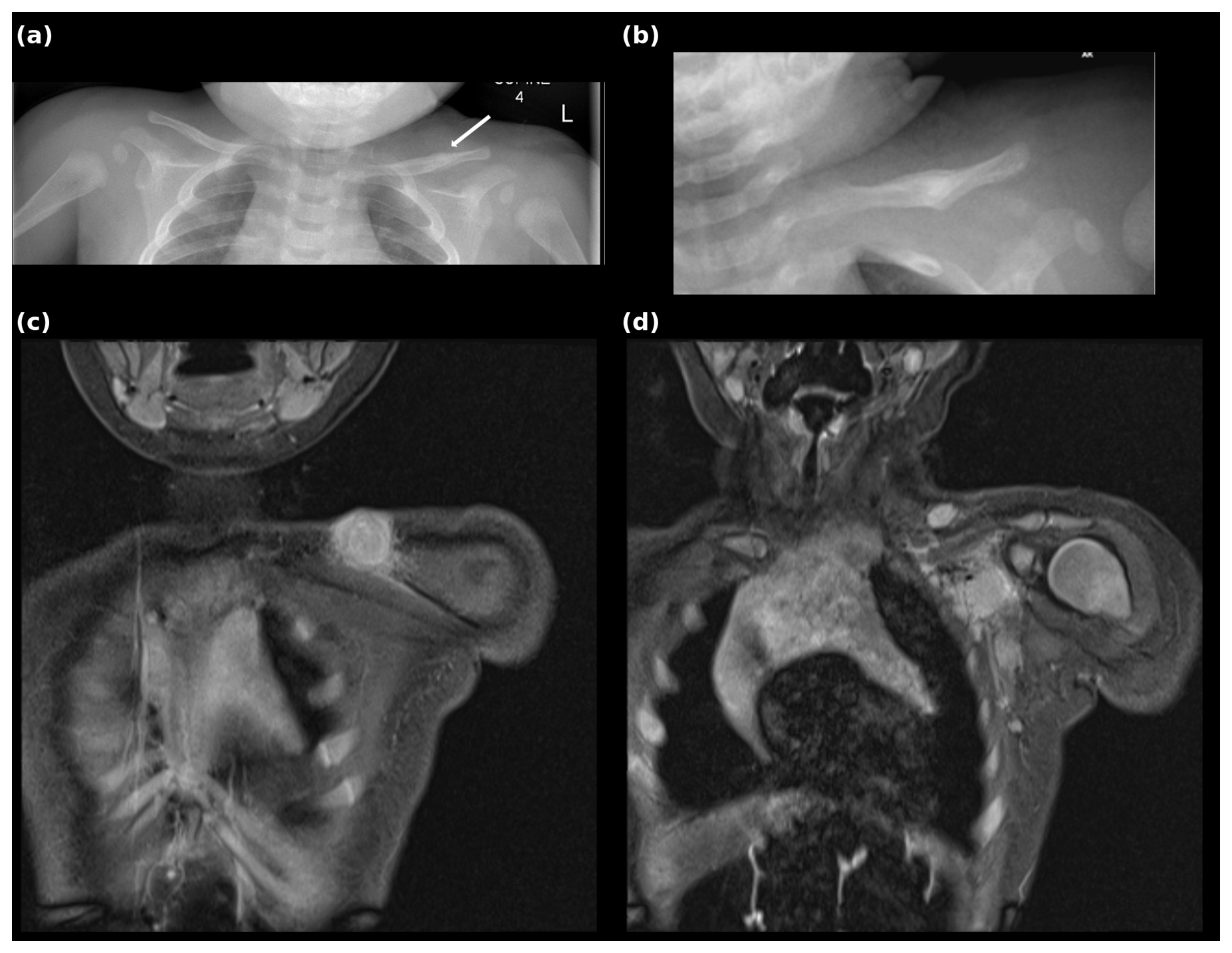
Figure 1Imaging of the left clavicle, shoulder, and axilla. (a, b) Anteroposterior radiograph of both clavicles 6 weeks after vaccination; the right clavicle is normal, while the arrow indicates periosteal reaction and periosteal reaction at the middle left clavicle. (c) Coronal magnetic resonance image showing a left supraclavicular collection of high proton density signal, measuring 1.5 cm × 1.9 cm, immediately superior and anterior to the middle third of the left clavicle. (d) A more posterior coronal slice showing enlarged left axillary lymph nodes.
By 8 weeks, repeat plain radiography showed a progressive callus, and ultrasound confirmed a complex echogenic inflammatory mass, a small anechoic collection at the injection site with axillary and supraclavicular lymphadenopathy. MRI revealed a 1.5 cm × 1.9 cm supraclavicular collection tracking from enlarged axillary nodes of up to 1.2 cm (Fig. 1c, d). A low T1 signal and circumferential clavicular cortical thickening were interpreted as secondary surface changes from adjacent infection rather than primary osteomyelitis. Manual aspiration yielded white purulent material culturing M. bovis BCG. Wild-type M. tuberculosis was excluded via PCR, and a comprehensive immunological screen comprising lymphocyte subsets, immunoglobulins, and phagocyte function was entirely normal.
A chest radiograph excluded pulmonary involvement. An electrocardiogram to establish a baseline corrected QT interval and baseline blood tests including liver function were obtained to monitor for treatment medication toxicity.
The child was treated with isoniazid, rifampicin, and moxifloxacin alongside pyridoxine supplementation for a planned total duration of 12 months. While using moxifloxacin in a newborn is off label and carries cartilage concerns, the clinical priority was the eradication of M. bovis and the prevention of central nervous system dissemination. Since M. bovis is intrinsically resistant to pyrazinamide, the low theoretical risk of serious arthropathy was considered to be an acceptable compromise for this defined treatment period. The infant responded well and continues to be monitored by the infectious-disease and orthopaedic teams. At this early stage, further radiographs are planned for 6 months to assess resolution.
BCG-related bone complications are traditionally attributed to haematogenous seeding, typically presenting 2 to 46 months post-vaccination at long-bone metaphyses (Lew and Waldvogel, 2004; Tsai et al., 2020). This case demonstrates a different pathophysiological mechanism with the unusually rapid 6-week onset (Alawfi, 2022; Lin et al., 2015). MRI confirmed that an injection site abscess tracked contiguously through axillary nodes to the supraclavicular area, causing secondary reactive surface changes on the adjacent clavicle rather than primary osteomyelitis.
Clavicular periosteal reaction lacking trauma history appropriately triggers NAI investigations. However, progressive soft tissue inflammation, spontaneous purulent discharge, and failure of standard anti-staphylococcal treatment must shift clinical suspicion toward atypical mycobacterial infection, particularly when the lesions arise adjacent to a recent vaccination site.
Severe or rapid post-vaccination complications require immunological evaluation to exclude underlying primary immunodeficiencies. (Al-Muhsen and Casanova, 2008; Bustamante et al., 2014). Localised BCG infection can arise in immunocompetent infants, as seen here. Diagnosis relies on the isolation of M. bovis BCG. Treatment necessitates prolonged antimycobacterial therapy tailored to its intrinsic pyrazinamide resistance (Ritz et al., 2009). Although international practices vary, including shorter antimicrobial regimens, our multidisciplinary team planned a 12-month course to ensure complete eradication and to safeguard against central nervous system (CNS) dissemination.
BCG-associated suppurative lymphadenitis with secondary reactive clavicular surface changes can develop early, within 6 weeks of vaccination, even in immunocompetent neonates. While unexplained infant clavicular periosteal reactions correctly prompt NAI investigations, atypical mycobacterial infection with contiguous spread must remain on the differential diagnosis when lesions occur near vaccination sites and resist standard antibiotic therapy.
The complete clinical data for this case report are not publicly available to protect the minor patient's privacy and to comply with UK data protection laws and institutional ethical guidelines.
PGN, RH, and AS provided clinical care for the patient. PGN, DF, SS, and RH collected and interpreted the clinical and imaging data. DF, SS, and PGN prepared the paper with contributions from all of the authors. AS supervised the work and obtained written informed consent from the patient's parents. All of the authors have read and approved the submitted version.
The contact author has declared that none of the authors has any competing interests.
This report describes the clinical care of a single patient. Written informed consent for publication of this case report, including the clinical details and the imaging presented in Fig. 1, was obtained from the patient's parents as his legal guardians; the signed consent documentation is held by the authors and is available to the editorial office on request. The patient is a minor. His identity and privacy have been protected throughout.
Publisher's note: Copernicus Publications remains neutral with regard to jurisdictional claims made in the text, published maps, institutional affiliations, or any other geographical representation in this paper. The authors bear the ultimate responsibility for providing appropriate place names. Views expressed in the text are those of the authors and do not necessarily reflect the views of the publisher.
Many thanks are given to Anish Sanghrajka for overseeing this project and the parents of patient for allowing this case report to be written up.
No generative artificial intelligence tool was used to prepare this paper.
This paper was edited by Derek Amanatullah and reviewed by two anonymous referees.
Alawfi, A.: Bacillus Calmette–Guérin Vaccine-Related Osteomyelitis in Immunocompetent Children in Saudi Arabia: A Narrative Review, Cureus, https://doi.org/10.7759/cureus.32762, 2022.
Al-Muhsen, S. and Casanova, J.-L.: The genetic heterogeneity of mendelian susceptibility to mycobacterial diseases, J. Allergy Clin. Immun., 122, 1043–1051, https://doi.org/10.1016/j.jaci.2008.10.037, 2008.
Bustamante, J., Boisson-Dupuis, S., Abel, L., and Casanova, J.-L.: Mendelian susceptibility to mycobacterial disease: Genetic, immunological, and clinical features of inborn errors of IFN-γ immunity, Semin. Immun., 26, 454–470, https://doi.org/10.1016/j.smim.2014.09.008, 2014.
Grange, J. M.: Complications of bacille Calmette–Guérin (BCG) vaccination and immunotherapy and their management, Commun. Dis. Public Health, 1, 84–88, 1998.
Kröger, L., Korppi, M., Brander, E., Kröger, H., Wasz-Höckert, O., Backman, A., Rapola, J., Launiala, K., and Katila, M.-L.: Osteitis Caused by Bacille Calmette–Guérin Vaccination: A Retrospective Analysis of 222 Cases, J. Infect. Dis., 172, 574–576, 1995.
Lew, D. P. and Waldvogel, F. A.: Osteomyelitis, Lancet, 364, 369–379, https://doi.org/10.1016/S0140-6736(04)16727-5, 2004.
Lin, W.-L., Chiu, N.-C., Lee, P.-H., Huang, A. S.-E., Huang, F.-Y., Chi, H., Huang, D. T.-N., and Chan, P.-C.: Management of Bacillus Calmette–Guérin osteomyelitis/osteitis in immunocompetent children – A systematic review, Vaccine, 33, 4391–4397, https://doi.org/10.1016/j.vaccine.2015.07.039, 2015.
Ritz, N., Tebruegge, M., Connell, T. G., Sievers, A., Robins-Browne, R., and Curtis, N.: Susceptibility of Mycobacterium bovis BCG vaccine strains to antituberculous antibiotics, Antimicrob. Agents Chemother., 53, 316–318, https://doi.org/10.1128/AAC.01302-08, 2009.
Teo, S. S. S. and Shingadia, D. V.: Does BCG have a role in tuberculosis control and prevention in the United Kingdom?, Archives of Disease in Childhood, 91, 529–531, https://doi.org/10.1136/adc.2005.085043, 2006.
Tsai, Y.-L., Chen, Y.-J., and Lai, Y.-C.: Long-term follow-up of osteomyelitis caused by Bacille Calmette–Guérin vaccination in immunocompetent children, Sci. Rep., 10, 4072, https://doi.org/10.1038/s41598-020-61152-6, 2020.
World Health Organization: BCG vaccine: WHO position paper, February 2018 – Recommendations, Vaccine, 36, 3408–3410, https://doi.org/10.1016/j.vaccine.2018.03.009, 2018.