the Creative Commons Attribution 4.0 License.
the Creative Commons Attribution 4.0 License.
 | 18 Nov 2026
Frequency and patterns of radiographic changes in chronic periprosthetic hip joint infection: a retrospective cohort study
Julius Michael Wolfgart

Moritz Mederake
Julie Hartmann
Sina Wiegand
Ulf Krister Hofmann
Bernd Fink
Conventional radiography remains a cornerstone in the diagnostics of periprosthetic complications, yet its ability to detect infection remains unclear. This study aimed to characterize the spectrum and frequency of radiographic changes in chronic periprosthetic hip joint infection (PJI) and to explore potential associations with underlying microbiological characteristics. A total of 133 patients with confirmed chronic periprosthetic hip joint infection undergoing prosthesis explantation were included. Standardized radiographs were systematically evaluated for characteristic signs, including periprosthetic radiolucent lines, focal osteolysis, heterotopic ossification, cortical hypertrophy, periosteal reaction, intracortical fistula formation, and periprosthetic fracture. These features were further analyzed for associations with the microbiological findings categorized by pathogen group, virulence, and polymicrobial infection in an exploratory fashion. Radiographic abnormalities were common, with 62.4 % of patients showing two or more characteristic signs (median 2, IQR 1–5). However, more than one-third of patients (37.6 %) exhibited either no abnormal findings or only one abnormal finding. The most frequent findings were radiolucent lines (55.6 %), heterotopic ossification (51.1 %), and cortical hypertrophy (49.2 %), followed by osteolysis (41.4 %), periosteal reaction (37.6 %), fracture (20.3 %), and sinus tract formation (7.5 %). Exploratory analyses indicated that polymicrobial infections were associated with a higher prevalence of acetabular radiolucent lines (14.8 % vs. 2.8 %, p = 0.031). Femoral osteolysis was more frequent in infections caused exclusively by Gram-positive pathogens (42.6 % vs. 0 %, p = 0.011). Given the small subgroups and wide confidence intervals, these findings are hypothesis-generating only. This study shows that plain radiographic abnormalities are common in chronic PJI. To our knowledge, a structured analysis of their frequencies and patterns has not been reported. Conventional radiography may provide relevant information on disease extent and chronicity, which could be of value in the clinical assessment of PJI, particularly given the diagnostic challenges of low-grade infections. However, as no aseptic control group was included, these patterns should be regarded as descriptive rather than diagnostic of chronic PJI. Comparing these results to aseptic cases in revision arthroplasty of the hip should be part of future investigations.
- Article
(2541 KB) - Full-text XML
- BibTeX
- EndNote


Periprosthetic joint infection (PJI) represents one of the most devastating complications in modern arthroplasty. In Germany, the annual number of primary total hip and knee arthroplasties increased by 5.4 % to more than 530 000 procedures in 2024 (Statistisches Bundesamt, 2024, 2025). Naturally, this increase is accompanied by a growing number of implant-related complications; during the same year, 60 922 revision procedures were performed at these two joints (Statistisches Bundesamt, 2025). The discrimination of an aseptic loosening from a PJI, however, remains an ongoing challenge (Alt et al., 2025). The cornerstones of diagnosis remain microbiological culture (McNally et al., 2021), while synovial leukocyte analysis is an important supportive – but not standalone – criterion (Sabater-Martos et al., 2025). Microbiological detection of isolates could be further improved by the sonication of prosthesis explants and was incorporated as a diagnostic criterion in the European Bone and Joint Infection definition (EBJIS) (McNally et al., 2021). Histopathological evaluation of the periprosthetic membrane has been an established diagnostic tool for decades (Morawietz et al., 2006; Krenn et al., 2014). In recent years, PCR (polymerase chain reaction)-based tissue analysis (Qu et al., 2013) and synovial fluid biomarkers such as leukocyte esterase (Poursalehian et al., 2025) and alpha-defensin (Abdelnasser et al., 2025) have been investigated as additional diagnostic tools. Additional biomarkers, including calprotectin (Festa et al., 2024), pentraxin-3 (Logroscino et al., 2023), and myeloperoxidase (Ikeda et al., 2025), are currently being further investigated.
Conventional radiography represents a fundamental imaging modality in the diagnostic workup of any complication following joint arthroplasty. Radiological signs of loosening within the first 5 years after implantation are listed among the clinical criteria for “infection likely” in the European Bone and Joint Infection Society (EBJIS) definition of PJI. However, the criteria specify neither the imaging modality nor the type of radiological findings, and the diagnostic value in this context remains poorly described (McNally et al., 2021). The radiopaque properties of bone, metallic implant components, and bone cement generate characteristic contrast patterns on plain radiographs that allow for the assessment of implant positioning, fixation, and structural integrity, as well as changes in the surrounding bone architecture (Fritz et al., 2013). In PJIs, bacterial colonization of the joint leads to enzymatic degradation processes affecting the periprosthetic tissue, most notably bone (Ormsby et al., 2021; Cassat et al., 2013). Described features in PJI thus include periosteal reaction, heterotopic ossification, intracortical sinus tract formation, focal osteolysis, pronounced soft tissue swelling compared with the contralateral side, radiolucency at the bone–implant interface, loosening of the implant, and periprosthetic fracture (Hofmann et al., 2024).
Beyond conventional radiography, a recent review and meta-analysis (Abedi et al., 2026) evaluated the diagnostic accuracy of cross-sectional and nuclear imaging modalities in PJI, including CT, MRI, white blood cell scintigraphy, FDG-PET/CT, antigranulocyte scintigraphy, and bone scintigraphy. Of note is that no eligible studies were included for CT and MRI. The overall diagnostic accuracy for the remaining modalities was found to be very heterogeneous, and no single technique was consistently superior, while white blood cell scintigraphy was described to perform most reliably when combined with bone or bone marrow scintigraphy.
Acute infections caused by highly virulent pathogens such as Staphylococcus aureus are typically more direct to diagnose and manage as they frequently present with pronounced local inflammatory signs and, often, systemic symptoms including fever and chills. During this relatively short time interval from onset of infection until treatment of a critically ill patient, usually, no radiographic changes of the bone around the implant become apparent. In contrast, low-virulence organisms such as Staphylococcus epidermidis or Cutibacterium acnes often do not lead to typical signs of infection such as redness, swelling, or fever. These low-grade infections are rather a chronic process developing over months or even years, potentially allowing for radiographic changes to develop. At the same time, these bacteria are often hard to detect by microbiological culture and, in some cases, an infection has to be assumed without ever being able to identify a causative germ (culture-negative PJIs). It is in these cases that characteristic radiographic changes might help the treating physician to decide upon or against an assumed low-grade infection and to initiate treatment accordingly. However, to date, the available literature on radiographic changes in PJIs includes only a very limited number of studies addressing this issue, often coming from a past era of arthroplasty, with small cohorts or short follow-up periods (reviewed in Hofmann et al., 2024). The establishment of a dedicated Imaging Working Group within the Unified PJI Definition Task Force further reflects the growing recognition of this diagnostic gap and the topic's clinical relevance (Abedi et al., 2026).
The aim of the present study was to characterize the frequency and patterns of radiographic findings in chronic PJI of the hip and to explore potential associations with the underlying microbiological spectrum.
2.1 Patients
The patients included in the present study were derived from a database of patients who underwent septic two-stage revision of a periprosthetic hip joint infection with implantation of a custom-made spacer, as previously described (Mederake et al., 2021, 2023). In total, 141 patients with late periprosthetic infection of the hip who underwent septic two-stage prosthesis revision surgery between April 2013 and October 2017 in the Orthopaedic Clinic Markgröningen were included. Six cases were excluded because microbiological cultures remained negative. Two cases were excluded due to missing radiographs. The patient cohort thus consisted of 133 cases with 56 (42 %) women and 77 (58 %) men, with a median age of 70 (27–92 years), as well as an average body mass index of 29.3±6.1. Diabetes mellitus was known in 24 patients (18 %), and a rheumatoid disease was known in 10 patients (7.5 %). Two patients were classified as ASA 1, 59 patients were classified as ASA 2, 69 patients were classified as ASA 3, and 3 patients were classified as ASA 4. The majority of explanted prostheses were cementless, followed by revision implants, as demonstrated in Table 1.
Table 1Types of infected prostheses. The table shows distribution of implant status and fixation type among the study population. The data are presented as absolute numbers and percentage of the total cohort. Hybrid: cementless cup with cemented stem. Reverse hybrid: cemented cup with cementless stem.
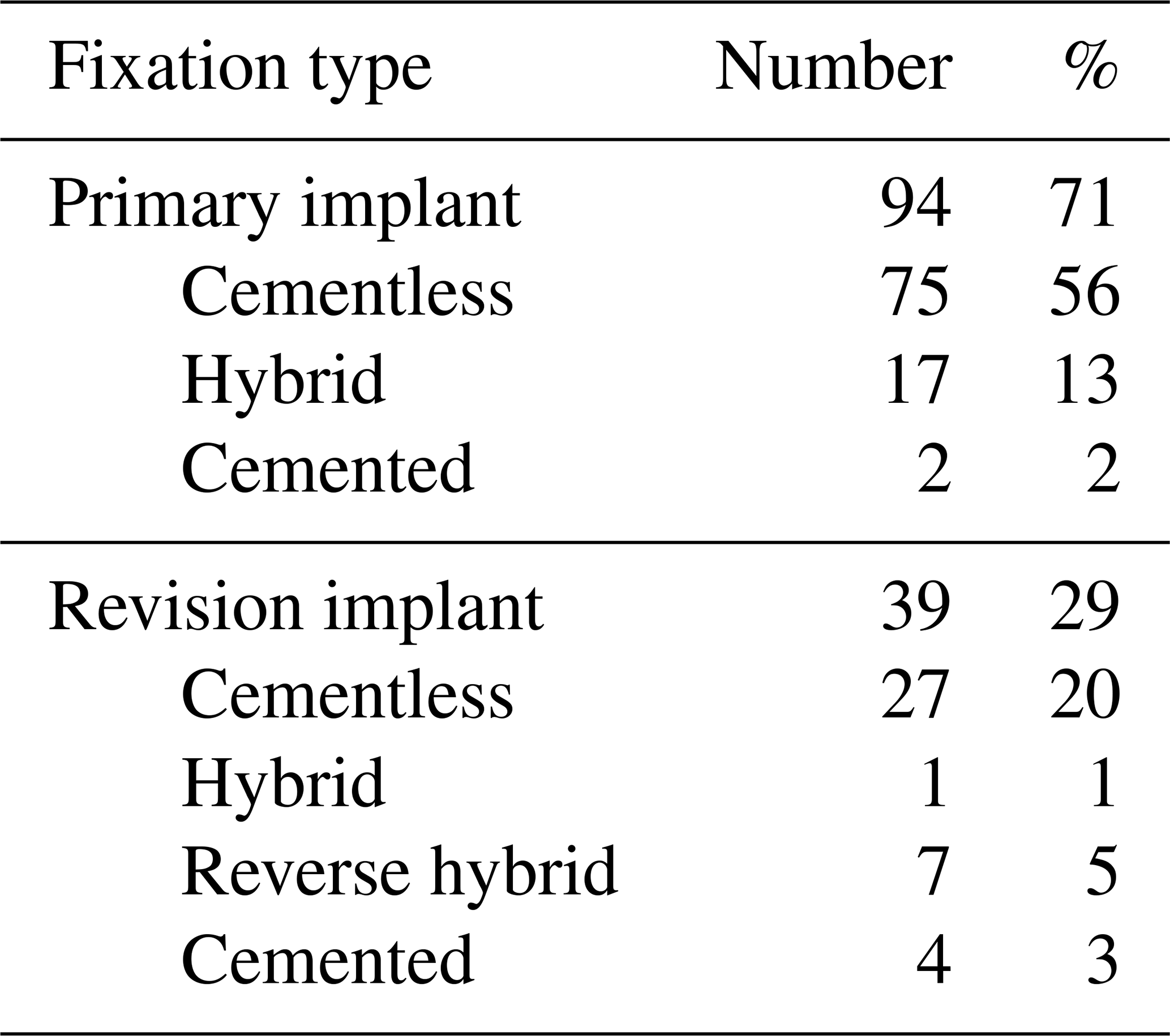
The periprosthetic joint infection was diagnosed preoperatively in all cases according to the criteria of the Musculoskeletal Infection Society (MSIS) (Parvizi et al., 2011). In 94 cases, a primary implant was involved, but there were also 39 patients who had already undergone at least one septic revision.
2.2 Methods
2.2.1 Microbiological diagnosis
A preoperative aspiration and/or biopsy with microbiological and histological examination of the hip joint was performed as standard procedure before any revision of a hip prosthesis. Bacteriological cultivation was assessed for 14 d according to Schäfer et al. (2008). Bacteriological and histological examination of the membrane at the site of loosening, which was removed during the operation, was carried out in accordance with the methods of Atkins et al. (1998), Virolainen et al. (2002), and Pandey et al. (1999) to confirm the original diagnosis. For statistical analysis, all detected pathogens were classified according to their virulence. High-virulence organisms included Staphylococcus aureus, Escherichia coli, Klebsiella pneumoniae, Klebsiella aerogenes, Proteus mirabilis, Morganella morganii, Streptococcus agalactiae, Enterococcus faecalis, Bacteroides fragilis, Candida albicans, Mycobacterium tuberculosis, Staphylococcus lugdunensis, Streptococcus sanguinis, Streptococcus mitis/oralis, Streptococcus salivarius, Streptococcus anginosus, Streptococcus gordonii, Granulicatella adiacens, and Listeria monocytogenes. Low-virulence organisms included Staphylococcus epidermidis, Staphylococcus hominis, Staphylococcus capitis, Staphylococcus haemolyticus, Staphylococcus saprophyticus, Staphylococcus warneri, Staphylococcus caprae, Cutibacterium acnes, Cutibacterium granulosum, Staphylococcus saccharolyticus, Actinomyces odontolyticus, and Lactobacillus plantarum. Pathogen virulence in PJI reflects a continuum rather than a dichotomous trait and is further influenced by host-related factors and the pathogen's capacity for biofilm formation, both of which were not accounted for in this noncomplex classification. Given the lack of an existing classification of pathogen virulence in PJI, the assignment was applied according to our own assessment, for exploratory purposes only, and should be interpreted with caution.
2.2.2 Radiographic evaluation
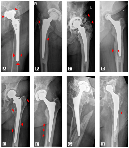
The parameters used for this study were based on a recent review article analyzing the diagnostic and prognostic relevance of plain radiographs for periprosthetic joint infections of the hip (Hofmann et al., 2024). The following parameters were thus registered and presented in Fig. 1: periosteal reaction, heterotopic bone formation, intracortical sinus tract formation, focal osteolysis, pronounced soft tissue collection compared to the contralateral side, periprosthetic fracture, implant loosening, or radiolucency around the implant. Pronounced soft tissue collection compared with the contralateral side and changes in implant position over time were not assessed. Following an initial screening of the radiographs, these parameters were excluded as pelvic overview images did not consistently display the entire soft tissue envelope around the hip, and prior imaging required for longitudinal assessment of implant position was not always available. One author (Julie Hartmann) performed the analysis of the radiographs. Uncertainties were put to discussion and mutually solved by a consensus of three authors (Julie Hartmann, Julius Michael Wolfgart, Ulf Krister Hofmann).
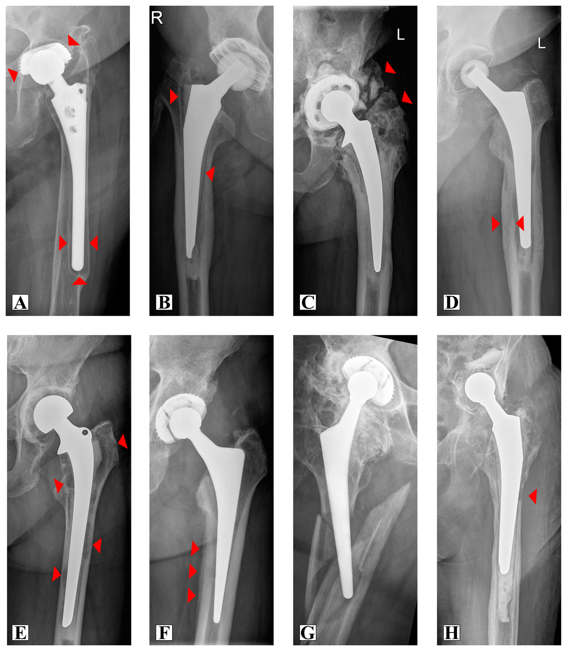
Figure 1Radiographic signs in chronic periprosthetic infection of the hip. Plain radiographs of the hip showing radiolucency at the bone–prosthesis interface (A), radiolucency at the bone–cement interface (B), heterotopic ossification (C), cortical hypertrophy (D), focal osteolysis (E), periosteal reaction (F), periprosthetic fracture of the femur (G), and intracortical sinus tract (H). Red arrows indicating corresponding features.
2.2.3 Statistical evaluation
Statistical analyses were performed using IBM SPSS Statistics Version 27 (IBM Corp., Armonk, NY, USA) and Microsoft Excel (Microsoft Corp., Redmond, WA, USA). Data distributions were assessed visually using histograms. Due to non-normal and skewed distributions, a non-parametric analytical approach was applied. Continuous variables are reported as medians with interquartile ranges or ranges, as appropriate, while categorical variables are presented as absolute frequencies and percentages. Associations between categorical variables were assessed using the χ2 test or Fisher's exact test when expected cell counts were low. Monte Carlo simulation was applied in cases of sparse contingency tables. Correlations were analyzed using Spearman's rank correlation coefficient. To account for polymicrobial infections, group-level variables were constructed to reflect the presence of specific microbial categories per case. All statistical tests were two-sided, with a predefined significance level of α = 0.05.
3.1 Radiographic changes
The number of positive radiographic signs per patient ranged from 0 to 7, with a median of 2 (IQR 1–5). At least one radiographic sign was present in 103 of 133 patients (77.4 %). Nearly half of the patients (47.4 %) demonstrated three or more radiographic signs. However, more than one-third of patients (37.6 %) exhibited either no abnormal findings or only one abnormal finding. The detailed frequency of radiographic signs is illustrated in Table 2.
Table 2Count of radiographic signs in plain radiography with chronic periprosthetic joint infections. The table illustrates the total number of characteristic signs (0–7) in plain radiography of the hip with chronic periprosthetic infection. The possible features included radiolucency at the bone–prosthesis interface, radiolucency at the bone–cement interface, heterotopic ossification, cortical hypertrophy, focal osteolysis, periosteal reaction, periprosthetic fracture of the femur, and intracortical sinus tract. The data are presented as absolute numbers, percentage of the total cohort, and cumulative percentages (rounded).
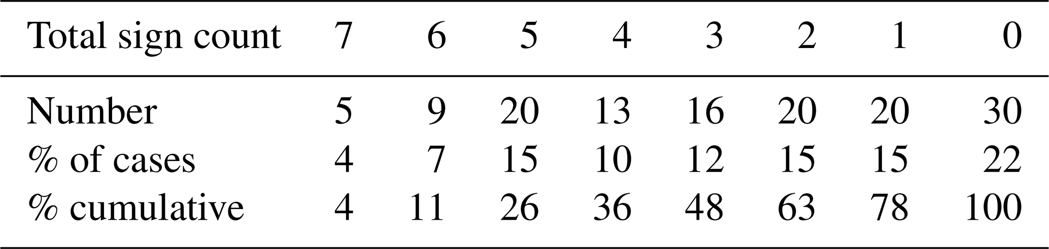
Radiolucent lines (55.6 %), heterotopic ossification (51.1 %), and cortical hypertrophy (49.2 %) were the most frequently observed radiographic signs in PJI. Radiolucency predominantly occurred at the bone–prosthesis interface (36.8 %), whereas involvement of the bone–cement interface was less common (20.9 %), and radiolucent lines at the cement–prosthesis interface were rare (4.5 %) and confined to the femoral stem. Focal osteolysis (41.4 %) and periosteal reaction (37.6 %) were commonly detected. Periprosthetic fractures occurred in 20.3 % of cases and were exclusively localized to the femur. Intracortical sinus tract formation was rare and only observed in 7.5 % of cases. A detailed count and distribution of radiographic signs is illustrated in Table 3.
Table 3Total number and proportion of plain radiographic signs with chronic periprosthetic joint infections of the hip (n=133). The table illustrates the total number and proportion of every specific feature within the study population. The proportion of radiolucency is only given for the corresponding fixation category.
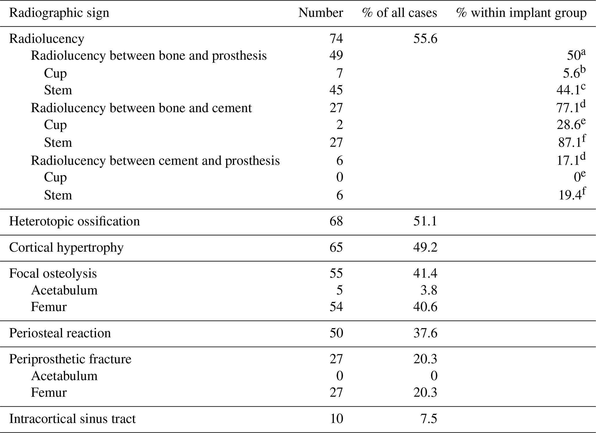
a Cementless THA (n=98), b cementless cup (n=126), c cementless stem (n=102), d cemented or hybrid (cementless cup, cemented stem) THA (n=35), e cemented cup (n=7), f cemented stem (n=31). THA – total hip arthroplasty.
3.2 Microbiological etiology
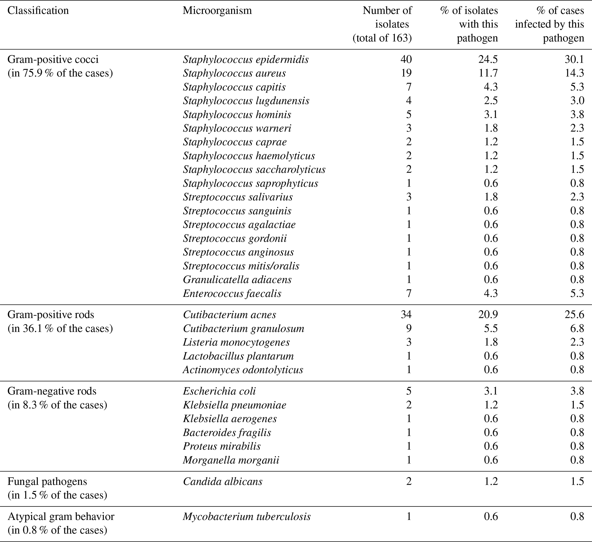
A polymicrobial infection with the simultaneous detection of at least two different microorganisms was observed in 27 patients (20.3 %), whereas monomicrobial infections were identified in 106 patients (79.7 %). The concurrent detection of exactly two different microorganisms occurred in 25 patients (18.8 %), while the simultaneous detection of three and four different microorganisms was observed in one patient each (0.75 %). Overall, 31 different microorganisms were identified from a total of 163 microbial isolates across the 133 cases (Table 4). The most frequently detected pathogen groups were Gram-positive cocci with 101 isolates (present in 75.9 % of cases), followed by Gram-positive rods with 48 isolates (36.1 % of cases), Gram-negative rods with 11 isolates (8.3 % of cases), fungi with two isolates (1.5 % of cases), and atypical pathogens with one isolate (0.8 % of cases). The three most frequently identified pathogens were Staphylococcus epidermidis, Cutibacterium acnes, and Staphylococcus aureus, with 40 (30.1 % of cases), 34 (25.6 % of cases), and 19 (14.3 % of cases) isolates, respectively. When stratified by virulence, 120 isolates (73.6 %) were classified as low-virulence pathogens, whereas 43 isolates (26.4 %) were classified as high-virulence pathogens. Low-virulence pathogens were detected in 89 (66.9 %) cases, whereas high-virulence pathogens were present in 51 (38.3 %) cases. Mixed infections with both low- and high-virulence pathogen types occurred in 7 (5.3 %) cases. Multidrug-resistant organisms were identified in six cases (4.5 %). The detailed frequency and distribution of microorganisms detected are illustrated in Table 4.
Table 4Identified microorganisms in cases of chronic periprosthetic infection of the hip. The table illustrates the total number and proportion of isolates (total of 163) for each microorganism detected along with the proportion of cases infected by the corresponding microorganism within the study population.
3.3 Exploratory analyses
3.3.1 Group comparisons
To investigate the influence of the microbiological spectrum on radiographic findings, several groups were defined: first septic revision (yes vs. no), virulence (only low virulence vs. only high virulence, mixed infections excluded), multidrug resistance (yes vs. no), polymicrobial infection (yes vs. no), coagulase characteristics (only coagulase-negative staphylococci vs. others), Gram characteristics (only Gram-positive pathogens vs. only Gram-negative pathogens or mixed Gram-negative Gram-positive pathogens). In addition, a radiographic score (0–1, 2–3, 4, 5–7 signs) was defined.
Polymicrobial infections were associated with a significantly higher prevalence of a radiolucent band between the implant and bone in the cup region compared with monomicrobial infections (, 14.8 %, 95 % CI 5.9 %–32.5 % vs. , 2.8 %, 95 % CI 1.0 %–8.0 %; p = 0.031). In addition, a significantly higher prevalence of focal osteolysis in the femoral stem region was observed in infections caused by Gram-positive pathogens compared with infections caused by only Gram-negative pathogens or mixed Gram-negative Gram-positive pathogens (, 42.6 %, 95 % CI 34.2 %–51.5 % vs. , 0 %, 95 % CI 0.0 %–29.9 %; p = 0.011).
There were no significant associations between the groups in terms of first septic revision, virulence, multidrug resistance or coagulase characteristics, and distinct radiographic signs were shown. Likewise, no significant influence of polymicrobial infection, Gram characteristics, coagulase characteristics, or virulence on the radiographic score could be demonstrated.
3.3.2 Correlation analyses
The time interval between primary implantation and explantation of the endoprosthesis and the radiographic score (0–1, 2–3, 4, 5–7 signs) showed a near-moderate positive correlation (ρ = 0.296, p<0.001). No relevant associations were found between the time interval from primary implantation to explantation and the presence of polymicrobial infection, antimicrobial resistance, or virulence.
The present study investigated the frequency and patterns of radiographic findings in chronic periprosthetic hip joint infection and analyzed associations with the underlying pathogen spectrum in an exploratory fashion. The results demonstrate that radiographic abnormalities are common in the setting of periprosthetic joint infections, which is in line with previous works on that topic (Bergström et al., 1974; Ure et al., 1998) and is consistent with the comprehensive overview of imaging approaches to PJI by Jardon et al. (2024). Two-thirds of the patients show two or more distinct radiologic features of infection. Nearly half of the patients with chronic PJI of the hip show multiple characteristic radiographic signs. While some of these characteristics seem to be characteristic of an infection such as a sinus tract, other findings may overlap and can be similar in both aseptic loosening and PJI. As no aseptic comparison group was included in the present study, the following comparisons are based exclusively on previously published literature. Radiolucent lines, for example, were observed in 55.6 % of cases but are also a well-established feature of both septic and aseptic implant failure. In cases of aseptic loosening, however, radiolucencies are typically attributed to fibrous tissue interposition caused by mechanical micromotion and progressive loss of osseointegration (Apostu et al., 2018). These changes usually evolve slowly over time and often appear to be relatively uniform and well demarcated along the implant interface (Wautier et al., 2020; He et al., 2025). In contrast, infectious loosening tends to progress more rapidly and is frequently associated with more aggressive bone destruction (Tigges et al., 1994; Temmerman et al., 2005), resulting from inflammation-driven osteolysis induced by bacterial biofilm formation and cytokine-mediated osteoclast activation (Josse et al., 2019). Consequently, infection-related radiolucent zones may present in a more irregular and extensive pattern. Focal osteolysis reflecting progressive bone destruction (Josse et al., 2019) was also observed in 41.4 % of cases in the present cohort. Comparable osteolytic patterns are described in aseptic implant failure as well, where stress shielding (Savio and Bagno, 2022) and wear-particle-induced bone resorption (Regis et al., 2019) frequently result in focal defects, for example, in the trochanteric region. Given the lack of comparative data, exact differences in frequency and pattern between those two entities remain unclear to date.
In the present study, focal osteolysis in the femoral stem region occurred more frequently in infections caused exclusively by Gram-positive pathogens. However, Gram staining reflects a microbiological classification based on cell wall structure rather than a direct marker of pathogen virulence, which makes interpretation of this observation challenging. Consequently, whether this association reflects pathogen-specific interactions with bone tissue or represents a coincidental finding cannot be determined from the present data. Ultimately, any biological explanation for this observation therefore remains speculative and would require validation in larger and independent cohorts.
Intracortical sinus tract formation may be interpreted as the most advanced stage of such focal osteolysis, representing complete cortical perforation caused by progressive bone destruction. In contrast to osteolytic defects, these findings are – to our knowledge – not observed in aseptic loosening and could thus be considered to be a specific trait for infection. Given the absence of a direct aseptic comparison group in the present study, this could not be formally verified. Sinus tract formation was, however, comparatively rare in the present cohort. In advanced stages of bone destruction, periprosthetic fractures may occur as the ultimate complication of progressive osteolysis and cortical breakdown. In such advanced stages of the disease, correct allocation of acute and pre-existing, iatrogenic fractures from previous revision surgeries can be difficult.
Heterotopic ossification represented the second most prevalent radiographic finding in the present cohort, occurring in more than half of all cases. Heterotopic ossifications are not exclusive to PJIs but may also occur in non-infected hip (Willburger et al., 2022) and knee (Rader et al., 1997) joints, including those without implanted endoprostheses (Meyers et al., 2019). The overall prevalence of heterotopic ossification after total hip arthroplasty is reported to be approximately 30 % (Zhu et al., 2015; Herzberg et al., 2024), while for the knee it is considerably lower, at 9 % (Rader et al., 1997). Prevalences for the occurrence of heterotopic ossifications in plain radiographs after septic revisions of hip arthroplasties ranged between 12 % and 84 % (Burastero et al., 2020; Manrique et al., 2018; Rosteius et al., 2019). Compared with aseptic revisions, however, septic cases showed a significantly higher prevalence of heterotopic ossification (84 % vs. 11 %) and, of note, higher Brooker grades (Manrique et al., 2018). A meta-analysis demonstrated a 2-fold-increased risk (RR 2.22; 95 % CI 1.00–4.91; p = 0.0497) for heterotopic ossification in PJIs of the hip (Pulik et al., 2025). These findings are supported by the data from the present study, in which heterotopic ossification was observed in 51.1 % of patients with chronic PJI of the hip, considerably exceeding the reported overall prevalence in cases without infection. Cortical hypertrophy likewise represents, at first glance, a seemingly unspecific characteristic following total hip arthroplasty. It is frequently observed in bicontact stems with diaphyseal fixation, where hypertrophy occurs at the level of stem anchorage, characterized by cortical thickening associated with increased structural density and preserved structural integrity (Kanto et al., 2020; Pulik et al., 2020). In these cases, however, cortical hypertrophy is mainly attributable to biomechanical adaptation (Yamako et al., 2014) in accordance with Wolff's law and, as such, presents as a thickened but highly intact cortical bone mostly at the site of cortical fixation (Kokoszka et al., 2015; Kanto et al., 2020). In contrast, infection-related cortical hypertrophy changes are predominantly initiated by molecular biological mechanisms (Massaccesi et al., 2022). Radiographic analysis in the present study demonstrated a diffuse and predominantly irregular and extensive cortical hypertrophy, with altered cortical bone architecture appearing to diverge from the typical biomechanically induced pattern. It has been suggested that infection of the bone by bacteria leads to paradoxical bone remodeling, involving concurrent appositional bone formation alongside loss of bone mass and structural integrity (Croes et al., 2019), which may result in an overall uneven and bulky picture. In earlier stages of the disease, this bone remodeling might be visible as a periosteal reaction preceding cortical hypertrophy. It reflects periosteal new bone formation and is generally interpreted as a response to any kind of periosteal stimulation due to, e.g., inflammation, tumor, or mechanical irritation. To date, two studies reported low sensitivity and moderate specificity of this feature for the diagnosis of periprosthetic joint infection (Lyons et al., 1985; Isern-Kebschull et al., 2020). In the present cohort, it was observed in 38 % of the cases.
Further analyses identified selected associations between radiographic findings and microbiological characteristics that should be interpreted in an exploratory, hypothesis-generating context, given the number of comparisons performed and the modest size of the underlying subgroups. Of note is that, for the association between polymicrobial infection and acetabular radiolucent lines, the 95 % confidence intervals of the two proportions showed slight overlap, highlighting the imprecision inherent to this small-subgroup comparison. In this context, periprosthetic radiolucent lines in the acetabular region of uncemented implants were observed significantly more frequently in polymicrobial infections, which are commonly associated with increased biofilm complexity and prolonged inflammatory activity rather than pathogen virulence alone (Costerton et al., 1999). Polymicrobial PJIs are commonly associated with delayed diagnosis and chronic disease courses, promoting progressive osteolysis at the bone–implant interface (Zimmerli et al., 2004; Tande and Patel, 2014). The acetabular region, characterized by extensive cancellous bone and a large implant–bone contact area, may therefore be particularly susceptible to these chronic osteolytic processes before becoming clinically symptomatic (Tigges et al., 1994). For context, implant survival time in the present cohort was prolonged, with a median interval of 86.8 months (IQR 22.9–167.3) between implantation and explantation. Given the wide confidence intervals, the clinical significance of these associations remains unclear, and they do not allow conclusions regarding pathogen-specific radiographic patterns.
Notably, more than one-third of patients (37.6 %) exhibited either no abnormal findings or only one abnormal finding on conventional radiographs. This suggests that normal radiographic findings of the hip cannot exclude chronic PJI and that conventional radiography has limited sensitivity for detecting an infection. Accordingly, at this point, radiographic findings should only be regarded as complementary to – not a substitute for – gold-standard diagnostic testing.
Overall, radiographic abnormalities are common in chronic PJI of the hip, but a relevant proportion of patients show no or only one pathological sign. These findings may help to characterize disease extent, but their diagnostic value remains to be established.
Study limitations
First, the retrospective design may introduce selection and information bias as only patients undergoing septic two-stage revision with available radiographs were included. Second, radiographic assessment was based on routine clinical images, which may vary in quality and projection, and interpretation of radiographic signs remains partly observer-dependent. Evaluation was performed by a single investigator, with consensus review by two more investigators in uncertain cases. However, formal interobserver agreement was not assessed, which should be considered when interpreting the reported findings. Third, the cohort consisted exclusively of patients with confirmed chronic periprosthetic hip joint infection, and no direct comparison with cases of aseptic loosening was performed. Consequently, several comparisons drawn throughout the discussion rely on previously published literature rather than on direct, formally tested comparisons within the present cohort, which limits conclusions regarding the specificity of individual radiographic signs for infection. In addition, including only culture-positive and two-stage chronic PJI cases subsequently leads to limited generalizability to the full clinical spectrum of chronic PJI. Finally, some subgroup analyses involved relatively small numbers of cases, particularly for cemented components or specific pathogen groups, and should therefore be interpreted cautiously and should be considered to be hypothesis-generating rather than confirmatory.
Plain radiographic structural changes are common in chronic PJI of the hip and predominantly reflect cumulative structural alterations over time rather than specific microbiological characteristics. However, more than one-third of patients had no abnormalities or only one, and so a normal radiograph does not rule out chronic PJI. To our knowledge, a systematic and structured analysis of the frequencies and patterns of radiographic changes in chronic periprosthetic hip joint infection has not yet been reported. The present study addresses this gap by providing a comprehensive characterization of these findings, which may help to assess disease extent and chronicity in patients with chronic PJI. Comparison with a control group undergoing aseptic revision surgery should be the focus of future investigations.
Due to data protection regulations and the terms of the ethical approval under which informed consent was obtained, the original dataset cannot be made publicly available. It can, however, be provided by the authors upon reasonable request, subject to institutional and ethical review.
JMW: conceptualization, methodology, formal analysis, data curation, visualization, writing (original draft, review and editing), project administration. MM: methodology, investigation, resources, writing (review and editing). JH: investigation, data curation, validation, writing (review and editing). SW: data curation, visualization, writing (review and editing). UKH: conceptualization, methodology, supervision, resources, writing (review and editing). BF: conceptualization, supervision, resources, writing (review and editing).
The contact author has declared that none of the authors has any competing interests.
This study was performed in line with the principles of the Declaration of Helsinki. Approval was granted by the Ethics Committee of the university hospital of Tübingen (registration no. 418/2021BO2).
Information is anonymized, and the submission does not include images that may identify persons. Informed consent was obtained from all individual participants included in the study.
Publisher's note: Copernicus Publications remains neutral with regard to jurisdictional claims made in the text, published maps, institutional affiliations, or any other geographical representation in this paper. The authors bear the ultimate responsibility for providing appropriate place names. Views expressed in the text are those of the authors and do not necessarily reflect the views of the publisher.
Initial language editing was conducted using Grammarly (Grammarly Inc., San Francisco, California, USA).
This open-access publication was funded by the RWTH Aachen University.
This paper was edited by Irene Katharina Sigmund and reviewed by two anonymous referees.
Abdelnasser, M. K., Bakhet, A., Hosni, A., Kamal, D. T., Osman, O. B., Abdelhameed, M. A., and Moustafa, M. M.: Alpha defensin immunoassay is more effective for ruling out rather than diagnosing periprosthetic joint infection (PJI): a prospective cohort study, Arthroplasty, 7, 52, https://doi.org/10.1186/s42836-025-00337-8, 2025.
Abedi, A. A., Tarabichi, S., Glaudemans, A. W. J. M., Parvizi, J., and the Imaging Working Group for the Unified PJI Definition Task Force: Imaging modalities demonstrate moderate and heterogeneous diagnostic accuracy for periprosthetic joint infection: A systematic review and meta-analysis, Knee Surg. Sport Tr. A., https://doi.org/10.1002/ksa.70423, 2026.
Alt, V., Szymski, D., Rupp, M., Fontalis, A., Vaznaisiene, D., Marais, L. C., Wagner, C., and Walter, N. on behalf of the Country Delegates of the European Bone and Joint Infection Society: The health-economic burden of hip and knee periprosthetic joint infections in Europe: a comprehensive analysis following primary arthroplasty, Bone Jt. Open, 6, 298–311, https://doi.org/10.1302/2633-1462.63.Bjo-2024-0225.R1, 2025.
Apostu, D., Lucaciu, O., Berce, C., Lucaciu, D., and Cosma, D.: Current methods of preventing aseptic loosening and improving osseointegration of titanium implants in cementless total hip arthroplasty: a review, J. Int. Med. Res., 46, 2104–2119, https://doi.org/10.1177/0300060517732697, 2018.
Atkins, B. L., Athanasou, N., Deeks, J. J., Crook, D. W., Simpson, H., Peto, T. E., McLardy-Smith, P., and Berendt, A. R.: Prospective evaluation of criteria for microbiological diagnosis of prosthetic-joint infection at revision arthroplasty. The OSIRIS Collaborative Study Group, J. Clin. Microbiol., 36, 2932–2939, https://doi.org/10.1128/JCM.36.10.2932-2939.1998, 1998.
Bergström, B., Lidgren, L., and Lindberg, L.: Radiographic Abnormalities Caused by Postoperative Infection Following Total Hip Arthroplasty, Clin. Orthop. Relat. R., 99, 95–102, 1974.
Burastero, G., Alessio-Mazzola, M., Cavagnaro, L., Chiarlone, F., Carrega, G., Capello, A. G., Lovisolo, S., and Felli, L.: Conservative two-stage revision with primary components of infected total hip arthroplasty: An analysis of survival, clinical and radiographic outcomes, PLoS One, 15, e0239981, https://doi.org/10.1371/journal.pone.0239981, 2020.
Cassat, J. E., Hammer, N. D., Campbell, J. P., Benson, M. A., Perrien, D. S., Mrak, L. N., Smeltzer, M. S., Torres, V. J., and Skaar, E. P.: A secreted bacterial protease tailors the Staphylococcus aureus virulence repertoire to modulate bone remodeling during osteomyelitis, Cell Host Microbe., 13, 759–772, https://doi.org/10.1016/j.chom.2013.05.003, 2013.
Costerton, J. W., Stewart, P. S., and Greenberg, E. P.: Bacterial Biofilms: A Common Cause of Persistent Infections, Science, 284, 1318–1322, https://doi.org/10.1126/science.284.5418.1318, 1999.
Croes, M., van der Wal, B. C. H., and Vogely, H. C.: Impact of Bacterial Infections on Osteogenesis: Evidence From In Vivo Studies, J. Orthop. Res., 37, 2067–2076, https://doi.org/10.1002/jor.24422, 2019.
Festa, E., Ascione, T., Di Gennaro, D., De Mauro, D., Mariconda, M., and Balato, G.: Synovial calprotectin in prosthetic joint infection. A systematic review and meta-analysis of the literature, Arch. Orthop. Traum. Su., 144, 5217–5227, https://doi.org/10.1007/s00402-024-05416-0, 2024.
Fritz, J., Lurie, B., and Miller, T. T.: Imaging of Hip Arthroplasty, Semin. Musculoskelet. Radiol., 17, 316–327, https://doi.org/10.1055/s-0033-1348098, 2013.
He, A., Zhang, Y., Lu, C., Cao, L., Liu, J., and Zhong, Z.: Imaging study of aseptic loosening of the acetabular cup after cementless total hip arthroplasty: a retrospective study, BMC Musculoskel. Dis., 26, 396, https://doi.org/10.1186/s12891-025-08607-x, 2025.
Herzberg, R., Tracey, O. C., Tahvilian, S., Baksh, N., Zikria, B., and Naziri, Q.: Incidence of heterotopic ossification following total hip arthroplasty by approach: a systematic review, Eur. J. Orthop. Surg. Tr., 34, 2089–2098, https://doi.org/10.1007/s00590-024-03896-9, 2024.
Hofmann, U., Eleftherakis, G., Migliorini, F., Fink, B., and Mederake, M.: Diagnostic and prognostic relevance of plain radiographs for periprosthetic joint infections of the hip: a literature review, Eur. J. Med. Res., 29, 314, https://doi.org/10.1186/s40001-024-01891-8, 2024.
Ikeda, S., Uchiyama, K., Minegishi, Y., Nakamura, M., Maehana, S., Eda, R., Kubo, M., Inoue, G., and Takaso, M.: Diagnostic Utility of a Rapid Myeloperoxidase Test in Synovial Fluid for Chronic Periprosthetic Joint Infection, J. Bone Joint Surg. Am., 107, 2574–2581, https://doi.org/10.2106/jbjs.24.01514, 2025.
Isern-Kebschull, J., Tomas, X., García-Díez, A. I., Morata, L., Moya, I., Ríos, J., and Soriano, A.: Value of multidetector computed tomography for the differentiation of delayed aseptic and septic complications after total hip arthroplasty, Skeletal Radiol., 49, 893–902, https://doi.org/10.1007/s00256-019-03355-1, 2020.
Jardon, M., Fritz, J., and Samim, M.: Imaging approach to prosthetic joint infection, Skeletal Radiol., 53, 2023–2037, https://doi.org/10.1007/s00256-023-04546-7, 2024.
Josse, J., Valour, F., Maali, Y., Diot, A., Batailler, C., Ferry, T., and Laurent, F.: Interaction Between Staphylococcal Biofilm and Bone: How Does the Presence of Biofilm Promote Prosthesis Loosening?, Front. Microbiol., 10, 1602, https://doi.org/10.3389/fmicb.2019.01602, 2019.
Kanto, M., Fukunishi, S., Fukui, T., Nishio, S., Fujihara, Y., Okahisa, S., Takeda, Y., Yoshiya, S., and Tachibana, T.: Radiological Evaluation of the Relationship Between Cortical Hypertrophy and Stress Shielding After Total Hip Arthroplasty Using a Cementless Stem, Arthroplast Today, 6, 894–900, https://doi.org/10.1016/j.artd.2020.09.018, 2020.
Kokoszka, P., Markuszewski, J., Łapaj, Ł., and Wierusz-Kozłowska, M.: Remodeling of bone tissue around the distal part of Zweymüller stem, Ortop. Traumatol. Rehabil., 17, 371–380, https://doi.org/10.5604/15093492.1173379, 2015.
Krenn, V., Morawietz, L., Perino, G., Kienapfel, H., Ascherl, R., Hassenpflug, G. J., Thomsen, M., Thomas, P., Huber, M., Kendoff, D., Baumhoer, D., Krukemeyer, M. G., Natu, S., Boettner, F., Zustin, J., Kölbel, B., Rüther, W., Kretzer, J. P., Tiemann, A., Trampuz, A., Frommelt, L., Tichilow, R., Söder, S., Müller, S., Parvizi, J., Illgner, U., and Gehrke, T.: Revised histopathological consensus classification of joint implant related pathology, Pathol. Res. Pract., 210, 779–786, https://doi.org/10.1016/j.prp.2014.09.017, 2014.
Logroscino, G., Fidanza, A., Giusti, I., Poppa, G., Troianello, L., Calafiore, F., Saracco, M., Avigni, R., Leone, R., Mantovani, A., Bottazzi, B., Grappiolo, G., and Loppini, M.: Pentraxin 3, a new biomarker for the diagnosis and management of PJI in primary and revision hip arthroplasty, Acta Biomed., 94, e2023100, https://doi.org/10.23750/abm.v94iS2.13770, 2023.
Lyons, C. W., Berquist, T. H., Lyons, J. C., Rand, J. A., and Brown, M. L.: Evaluation of radiographic findings in painful hip arthroplasties, Clin. Orthop. Relat. R., 195, 239–251, 1985.
Manrique, J., Alijanipour, P., Heller, S., Dove, M., and Parvizi, J.: Increased Risk of Heterotopic Ossification Following Revision Hip Arthroplasty for Periprosthetic Joint Infection, Arch. Bone Jt. Surg., 6, 486–491, 2018.
Massaccesi, L., Galliera, E., Pellegrini, A., Banfi, G., and Corsi Romanelli, M. M.: Osteomyelitis, Oxidative Stress and Related Biomarkers, Antioxidants (Basel), 11, https://doi.org/10.3390/antiox11061061, 2022.
McNally, M., Sousa, R., Wouthuyzen-Bakker, M., Chen, A. F., Soriano, A., Vogely, H. C., Clauss, M., Higuera, C. A., and Trebše, R.: The EBJIS definition of periprosthetic joint infection, Bone Joint J., 103-b, 18–25, https://doi.org/10.1302/0301-620x.103b1.Bjj-2020-1381.R1, 2021.
Mederake, M., Hofmann, U. K., and Fink, B.: New Technique for Custom-Made Spacers in Septic Two-Stage Revision of Total Hip Arthroplasties, Antibiotics, https://doi.org/10.3390/antibiotics10091073, 2021.
Mederake, M., Hofmann, U. K., and Fink, B.: Clinical evaluation of a new technique for custom-made spacers in septic two-stage revision of total hip arthroplasties, Arch. Orthop. Traum. Su., 143, 5395–5403, https://doi.org/10.1007/s00402-022-04748-z, 2023.
Meyers, C., Lisiecki, J., Miller, S., Levin, A., Fayad, L., Ding, C., Sono, T., McCarthy, E., Levi, B., and James, A. W.: Heterotopic Ossification: A Comprehensive Review, JBMR Plus, 3, e10172, https://doi.org/10.1002/jbm4.10172, 2019.
Morawietz, L., Classen, R. A., Schröder, J. H., Dynybil, C., Perka, C., Skwara, A., Neidel, J., Gehrke, T., Frommelt, L., Hansen, T., Otto, M., Barden, B., Aigner, T., Stiehl, P., Schubert, T., Meyer-Scholten, C., König, A., Ströbel, P., Rader, C. P., Kirschner, S., Lintner, F., Rüther, W., Bos, I., Hendrich, C., Kriegsmann, J., and Krenn, V.: Proposal for a histopathological consensus classification of the periprosthetic interface membrane, J. Clin. Pathol., 59, 591–597, https://doi.org/10.1136/jcp.2005.027458, 2006.
Ormsby, R. T., Zelmer, A. R., Yang, D., Gunn, N. J., Starczak, Y., Kidd, S. P., Nelson, R., Solomon, L. B., and Atkins, G. J.: Evidence for osteocyte-mediated bone-matrix degradation associated with periprosthetic joint infection (PJI), Eur. Cell Mater., 42, 264–280, https://doi.org/10.22203/eCM.v042a19, 2021.
Pandey, R., Drakoulakis, E., and Athanasou, N. A.: An assessment of the histological criteria used to diagnose infection in hip revision arthroplasty tissues, J. Clin. Pathol., 52, 118–123, https://doi.org/10.1136/jcp.52.2.118, 1999.
Parvizi, J., Zmistowski, B., Berbari, E. F., Bauer, T. W., Springer, B. D., Della Valle, C. J., Garvin, K. L., Mont, M. A., Wongworawat, M. D., and Zalavras, C. G.: New definition for periprosthetic joint infection: from the Workgroup of the Musculoskeletal Infection Society, Clin. Orthop. Relat. R., 469, 2992–2994, https://doi.org/10.1007/s11999-011-2102-9, 2011.
Poursalehian, M., Soltani Farsani, A., Tabatabaei Irani, P., Ayati Firoozabadi, M., Parvizi, J., and Mortazavi, S. J.: What is the best cutoff point of leukocyte esterase for diagnosis of periprosthetic joint infections? a systematic review and meta-analysis, Arthroplasty, 7, 41, https://doi.org/10.1186/s42836-025-00325-y, 2025.
Pulik, Ł., Mierzejewski, B., Ciemerych, M. A., Brzóska, E., and Łęgosz, P.: The Survey of Cells Responsible for Heterotopic Ossification Development in Skeletal Muscles – Human and Mouse Models, Cells, https://doi.org/10.3390/cells9061324, 2020.
Pulik, Ł., Łęgosz, P., Brzóska, E., Mierzejewski, B., Grabowska, I., Ciemerych, M. A., and Hube, R.: Periprosthetic joint infection and heterotopic ossification after total hip arthroplasty: systematic review and meta-analysis, BMC Musculoskelet. Di., 26, 768, https://doi.org/10.1186/s12891-025-09045-5, 2025.
Qu, X., Zhai, Z., Li, H., Li, H., Liu, X., Zhu, Z., Wang, Y., Liu, G., and Dai, K.: PCR-based diagnosis of prosthetic joint infection, J. Clin. Microbiol., 51, 2742–2746, https://doi.org/10.1128/jcm.00657-13, 2013.
Rader, C. P., Barthel, T., Haase, M., Scheidler, M., and Eulert, J.: Heterotopic ossification after total knee arthroplasty. 54/615 cases after 1–6 years' follow-up, Acta Orthop. Scand., 68, 46–50, https://doi.org/10.3109/17453679709003974, 1997.
Regis, D., Sandri, A., Samaila, E., and Magnan, B.: Two-stage management of a spontaneous fracture of the greater trochanter through osteolytic lesions induced by polyethylene wear of a total hip arthroplasty. A case report, Acta Biomed., 90, 177–182, https://doi.org/10.23750/abm.v90i1-S.8086, 2019.
Rosteius, T., Rausch, V., Pätzholz, S., Lotzien, S., Baecker, H., Schildhauer, T. A., and Geßmann, J.: Incidence and risk factors for heterotopic ossification following periprosthetic joint infection of the hip, Arch. Orthop. Traum. Su., 139, 1307–1314, https://doi.org/10.1007/s00402-019-03215-6, 2019.
Sabater-Martos, M., Clauss, M., Ribau, A., Sousa, R., and on behalf of the Leukocyte Count Synovial Fluid working group for the Unified PJI definition task force: Differential synovial fluid white blood cell count for the diagnosis of chronic peri-prosthetic joint infection – a systematic review and meta-analysis, J. Bone Joint Infect., 10, 165–184, https://doi.org/10.5194/jbji-10-165-2025, 2025.
Savio, D. and Bagno, A.: When the Total Hip Replacement Fails: A Review on the Stress-Shielding Effect, Processes, https://doi.org/10.3390/pr10030612, 2022.
Schäfer, P., Fink, B., Sandow, D., Margull, A., Berger, I., and Frommelt, L.: Prolonged bacterial culture to identify late periprosthetic joint infection: a promising strategy, Clin. Infect. Dis., 47, 1403–1409, https://doi.org/10.1086/592973, 2008.
Statistisches Bundesamt: Fallpauschalenbezogene Krankenhausstatistik (DRG-Statistik): Operationen und Prozeduren der vollstationären Patientinnen und Patienten in Krankenhäusern (4-Steller) 2023, Statistisches Bundesamt, 24 September 2024, WiesbadenEVAS-Nr. 23141, 2024.
Statistisches Bundesamt: Fallpauschalenbezogene Krankenhausstatistik (DRG-Statistik): Operationen und Prozeduren der vollstationären Patientinnen und Patienten in Krankenhäusern (4-Steller) 2024, 30 September 2025, Statistisches Bundesamt, WiesbadenEVAS-Nr. 23141, 2025.
Tande, A. J. and Patel, R.: Prosthetic joint infection, Clin. Microbiol. Rev., 27, 302–345, https://doi.org/10.1128/cmr.00111-13, 2014.
Temmerman, O. P. P., Raijmakers, P. G. H. M., Berkhof, J., Hoekstra, O. S., Teule, G. J. J., and Heyligers, I. C.: Accuracy of diagnostic imaging techniques in the diagnosis of aseptic loosening of the femoral component of a hip prosthesis, J. Bone Joint Surg. Br., 87-B, 781–785, https://doi.org/10.1302/0301-620X.87B6.15625, 2005.
Tigges, S., Stiles, R. G., and Roberson, J. R.: Appearance of septic hip prostheses on plain radiographs, AJR Am. J. Roentgenol., 163, 377–380, https://doi.org/10.2214/ajr.163.2.8037035, 1994.
Ure, K. J., Amstutz, H. C., Nasser, S. A. M., and Schmalzried, T. P.: Direct-Exchange Arthroplasty for the Treatment of Infection after Total Hip Replacement. An Average Ten-Year Follow-up*, J. Bone Joint Surg., 80, 961–968, 1998.
Virolainen, P., Lahteenmaki, H., Hiltunen, A., Sipola, E., Meurman, O., and Nelimarkka, O.: The reliability of diagnosis of infection during revision arthroplasties, Scand. J. Surg., 91, 178–181, https://doi.org/10.1177/145749690209100208, 2002.
Wautier, D., Ftaïta, S., and Thienpont, E.: Radiolucent lines around knee arthroplasty components: a narrative review, Acta Orthop. Belg., 86, 82–94, 2020.
Willburger, R. E., Brinkhoff, F., Nottenkämper, J., Krapp, J., and Oberberg, S.: Heterotopic ossification after total hip arthroplasty: When is development completed?, J. Orthop. Surg. Res., 17, 147, https://doi.org/10.1186/s13018-022-02959-z, 2022.
Yamako, G., Chosa, E., Zhao, X., Totoribe, K., Watanabe, S., Sakamoto, T., and Nakane, N.: Load-transfer analysis after insertion of cementless anatomical femoral stem using pre- and post-operative CT images based patient-specific finite element analysis, Med. Eng. Phys., 36, 694–700, https://doi.org/10.1016/j.medengphy.2014.02.018, 2014.
Zhu, Y., Zhang, F., Chen, W., Zhang, Q., Liu, S., and Zhang, Y.: Incidence and risk factors for heterotopic ossification after total hip arthroplasty: a meta-analysis, Arch. Orthop. Traum. Su., 135, 1307–1314, https://doi.org/10.1007/s00402-015-2277-8, 2015.
Zimmerli, W., Trampuz, A., and Ochsner, P. E.: Prosthetic-joint infections, N. Engl. J. Med., 351, 1645–1654, https://doi.org/10.1056/NEJMra040181, 2004.